Short Communication - Imaging in Medicine (2015) Volume 7, Issue 2
A novel linear accelerator based stereotactic radiosurgery system
Ning Wen*Department of Physics, Clinical Director,Henry Ford Health System, Detroit, MI, United States
- Corresponding Author:
- Ning Wen
Department of Physics
Clinical Director,Henry Ford Health System
Detroit, MI, United States
Tel: 1-248-661-6150
E-mail: nwen1@hfhs.org
Abstract
A novel platform for LINAC-based SRS treatment, the Edge, (Varian Medical Systems, Palo Alto, CA) offers multiple imaging modalities for treatment localization, including an optical surface monitoring system (OSMS) for surface tracking, 2.5 MV portal imaging, triggered monoscopic kV imaging to track intra-fractional motion, 4D CBCT to evaluate tumor motion offline, extended CBCT images by stitching multiple CBCT scans together, and a Calypso/Varian electromagnetic beacon-based tracking system. The new PerfectPitchTM couch supports six degrees-of-freedom (6DoF) corrections from multiple imaging modalities used for precise patient setup. The new flat panel imager (DMI) has a greater dynamic range and faster image readout rate to process high dose rate of flatten filter free beams without saturation.
Keywords
Stereotactic radiosurgery; edge; image guided radiation therapy; stereotactic
Body radiation therapy
There have been many breakthroughs in the technological development of the stereotactic radiosurgery (SRS) since 1951 [1-4]. The Linac based radiosurgery has been widely adopted after the accuracy has been significantly improved in 1980s. The multi-leaf collimators (MLC) have gradually replaced traditional conical cones as the leaves get finer to achieve similar dose conformity. Various treatment delivery techniques have been implemented for improve conformity to complex geometric targets and minimize the dose spread to normal tissues, such as dynamic arc delivery, Intensity Modulated Radiation Therapy (IMRT) and Volumetric Modulated Arc Therapy (VMAT) [5-7].
A novel platform for LINAC-based SRS treatment, the Edge, (Varian Medical Systems, Palo Alto, CA) offers multiple imaging modalities for treatment localization, including an optical surface monitoring system (OSMS) for surface tracking, 2.5 MV portal imaging, triggered monoscopic kV imaging to track intra-fractional motion, 4D CBCT to evaluate tumor motion offline, extended CBCT images by stitching multiple CBCT scans together, and a Calypso/Varian electromagnetic beaconbased tracking system. The new PerfectPitchTM couch supports six degrees-of-freedom (6DoF) corrections from multiple imaging modalities used for precise patient setup. The new flat panel imager (DMI) has a greater dynamic range and faster image readout rate to process high dose rate of flatten filter free beams without saturation.
Since the system was installed in March 2014, three manuscripts were published regarding the evaluation and implementation of the system from our group at Henry Ford Health System.
The manuscript titled “Characteristics of a novel treatment system for Linear Accelerator– based stereotactic radiosurgery”, accepted by the Journal of Applied Clinical Medical Physics, was the first paper published regarding the Edge system [8]. The study comprehensively characterized the dosimetric properties and imaging modalities of this treatment platform for localizing and treating patients with frameless, image guided stereotactic radiosurgery (SRS) and stereotactic body radiotherapy (SBRT). It was a multidisplinary collaboration with co-authors from the department of Radiation Oncology, Interventional Gastroenterology, Thoracic Surgery, Intervention Pulmonology and Neurosurgery. The Edge radiosurgery system has been shown to possess the high accuracy localization and delivery requirements for safely treating patients with tumors in the CNS, Spine, Lung, Liver, Pancreas, and Head & Neck in one to five fractions using SRS/ SBRTbased techniques.
The second manuscript titled “Targeting Accuracy of Image-Guided Radiosurgery for Intracranial Lesions: A Comparison across Multiple Linear Accelerator Platforms”, accepted by the Technology in Cancer Research & Treatment, quantitatively evaluated multiple Linear accelerator based system for intracranial SRS [9]. The Edge system was shown to have superior setup accuracy with the setup error of 0.3 ± 0.1 mm. The system has been proven to be efficient and accurate for intracranial SRS treatment using a non-invasive approach.
The third manuscript titled “Generation and verification of QFix kVue Calypso-compatible couch top model for a dedicated stereotactic linear accelerator with FFF beams”, accepted by the Journal of Applied Clinical Medical Physics, detailed the creation, verification, and implementation of a robotic couch used in the Edge system [10]. The new robotics couch supports six degrees-of-freedom corrections from multiple imaging modalities for precise patient setup. Our study developed an innovative approach to incorporate the couch model in the treatment planning system to ensure the precise dose delivery.
Frameless image-guided intracranial SRS has been emerging to treat brain metastasis or abnormalities. A stereotactic thermal plastic mask system is used to immobilize the patient’s head. The inter- and intra- fractional motion is monitored by x-ray or other imaging modalities. Successful treatment should be attributed to the highly rigid setup and therefore limiting intrafraction motion during treatment. For motion management, the Edge linear accelerator treatment platform (Varian Medical Systems), in addition to planar and volumetric imaging modalities, includes the OSMS, which provides real-time surface motion information and beam hold functionality. Recently, Encompass™ Intracranial Immobilization System, a novel stereotactic open mask system, has been developed by QFix as shown in FIGURE 1. This mask has an anterior open view around the eyes and nose through which the optical camerabased OSMS can monitor the patient motion in real-time. We investigated the capability of monitoring intrafractional motion of the patient within this mask coupled with OSMS.

Figure 1: Intracranial Immobilization System. (A) and (B) top and bottom pieces, (C) built-in head frame on the couch top, (D) fabricated bottom piece on the head frame, and (E) top piece affixed to the head frame. Built-in adjustable shims ranging from 0 to 4 mm and push-down locking pins at 6 points (indicated by arrow).
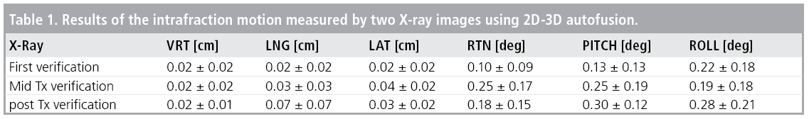
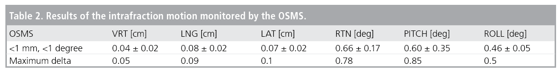
Intrafractional motions (6 dimension, translations and rotations) measured using two x-ray images were within 1 mm and 1 degree for all six patients (TABLE 1). Accuracy of OSMS monitoring depended on the selected ROI (TABLE 2). The OSMS monitored motion within 1 mm and 1° when the open surface area captured by the camera was greater than 33 cm2. When such condition was not met, the accuracy could be degraded.
In our studies, we systematically investigated the accuracy of a new generation of frameless, image guided radiosurgery platform and tested multiple aspects of the platform. The accuracy was demonstrated to be comparable to invasive frame-based and frameless based SRS systems with multiple imaging modalities.
References
- Leksell L. Sterotaxic radiosurgery in trigeminal neuralgia. Acta. Chir. Scand. 137, 311-314 (1971).
- Adler JR Jr, Chang SD, Murphy MJ et al. The Cyberknife: a frameless robotic system for radiosurgery. Stereotact. Funct. Neurosurg. 69, 124-128 (1997).
- Ryu S, Fang Yin F, Rock J et al. Image-guided and intensity-modulated radiosurgery for patients with spinal metastasis. Cancer. 97, 2013-2018 (2003).
- Garcia-Barros M, Paris F, Cordon-Cardo C et al. Tumor response to radiotherapy regulated by endothelial cell apoptosis. Science. 300, 1155-1159 (2003).
- Solberg TD, Boedeker KL, Fogg R et al. Dynamic arc radiosurgery field shaping: a comparison with static field conformal and noncoplanar circular arcs. Int. J. Radiat. Oncol. Biol. Phys. 49, 1481-1491 (2001).
- Jin JY, Chen Q, Jin R et al. “Technical and clinical experience with spine radiosurgery: a new technology for management of localized spine metastases. Technol. Cancer. Res. Treat. 6, 127-133 (2007).
- Audet C1, Poffenbarger BA, Chang P et al. Evaluation of volumetric modulated arc therapy for cranial radiosurgery using multiple noncoplanar arcs. Med. Phys. 38, 5863-5872 (2011).
- N Wen, H Li, K Song et al. “Characteristics of a novel treatment system for linear acceleratorbased stereotactic radiosurgery.” J. Appl. Clin. Med. Phys. 16, 5313-5319 (2015).
- Y Huang, B Zhao, IJ Chetty et al. “Targeting Accuracy of Image-Guided Radiosurgery for Intracranial Lesions: A Comparison Across Multiple Linear Accelerator Platforms.” Technol. Cancer. Res. Treat. (2015).
- Gardner SJ, Gulam M, Song K et al. Generation and verification of QFix kVue Calypso-compatible couch top model for a dedicated stereotactic linear accelerator with FFF beams. J. Appl. Clin. Med. Phys. 16, 5441 (2015).


