Review Article - Imaging in Medicine (2015) Volume 7, Issue 2
A review on single coronary artery
Abhisekh Mohanty*Department of Cardiology, Continental Hospitals, Hyderabad, India
- Corresponding Author:
- Abhisekh Mohanty
Department of Cardiology
Continental Hospitals, Hyderabad, India
Tel: +91 40 6700 0000
E-mail: abhisekh.mhnt@gmail.com
Abstract
Coronary artery anomalies are rare congenital disorders. They are usually diagnosed incidentally during coronary artery angiogram or post mortem examinations. Single coronary artery (SCA) anomalies are a subset of coronary artery anomalies. The prevalence of single coronary anomalies in the general population is approximately 0.024% according to Lipton’s reports. The various patterns of SCA are difficult to understand. Lipton et al. systematically classified the different types of SCA. Most of the cases of SCA are asymptomatic .The malignant variety of SCA is the type in which the coronary artery courses between the great vessels (Aorta and Pulmonary artery).
Keywords
Single coronary artery; lipton’s classification; anomalous coronary artery
Compression Coronary artery anomalies are usually diagnosed incidentally during coronary artery angiogram and post-mortem evaluation. The prevalence of coronary artery anomalies is less than 0.024–0.044% based on published articles [1].The most common coronary artery anomaly seen is the separate origin of the Left anterior descending (LAD) and Left circumflex (LCX) artery. The second most common anomaly detected is origin of LCX artery from Right coronary artery (RCA). The cases which will be discussed in this article are single coronary artery anomalies (SCA); these cases are rare, occurring in approximately 0.024% of the population according to Lipton’s reports. The various patterns of SCA are difficult to understand.
Single Coronary artery has been known since 1903. It is an anomalous condition in which the single coronary artery originates from either the left or right aortic sinus and gives rise to the entire coronary circulation. They may or may not be associated with congenital heart disease. A single coronary artery which is associated with structurally normal heart, usually divides into normally formed and normally distributed branches. Those cases which are associated with congenital heart diseases have abnormal branching patterns [2,3]. In a large series of 126,595 patients, 0.019% of patients had single coronary artery. Of these 40% were associated with congenital heart diseases like tetralogy of ballot, truncus arteriosus and transposition of great arteries [2]. The current classification of single coronary artery was proposed by Lipton et al. [1] (TABLE 1). Group I has anatomical course of either a right or left coronary artery. Group II anomalies arise from the proximal part of the normal right or left coronary artery, and cross the base of the heart before assuming the normal position of the inherent coronary artery. Group III describes the anomaly where the LAD artery and LCX artery arise separately from the proximal part of the normal RCA. Five anatomical subtypes exist, and are classified according to the relationship of the anomalous coronary artery with the aorta and pulmonary artery, i.e., “anterior,” “between,” “septal,” “posterior,” or “combined.” In this series, the “septal” subtype was the most common, whereas the “between” subtype was the least common (FIGURE 1).
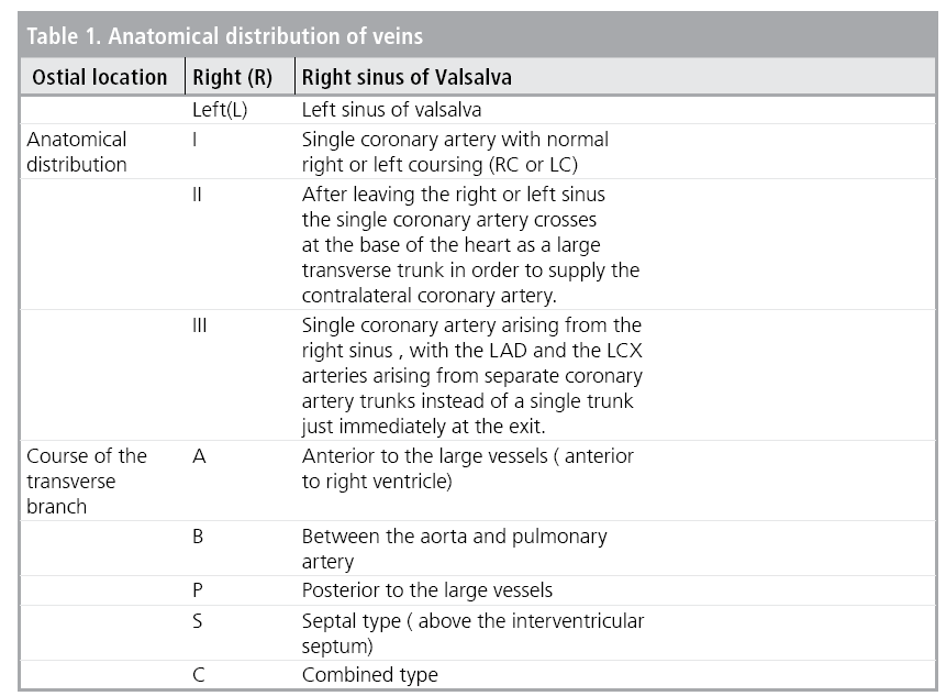

Figure 1: Lipton ’s classification of SCA.
Type R-I is the least common type of SCA. I had the privilege of diagnosing a case Lipton’s Type R-I SCA (FIGURE 2). Patients with Single coronary artery are usually asymptomatic. Sometimes they present with angina, syncope or sudden cardiac arrest. Myocardial infarction or sudden cardiac arrest may occur in cases in which the course of the coronary artery is in between the aorta and pulmonary artery. In such patients even though there is no treatment strategy, symptomatic patients can go for coronary artery bypass grafting or transposition of coronary arteries to appropriate coronary sinus [4].

Figure 2: Type R-I SCA.
References
- Yamanaka O, Hobbs RE. Coronary artery anomalies in 126,595 patients undergoing coronary arteriography. Cathet. Cardiovasc. Diagn. 21, 28-40 (1990).
- Roberts WC. Major anomalies of coronary arterial origin seen in adulthood. Am. Heart. J. 111, 941-963 (1986).
- Braun MU, Stolte D, Rauwolf T et al. Single coronary artery with anomalous origin from the right sinus Valsalva. Clin. Res. Cardiol. 95, 119-121 (2006).
- Mohanty A, Chandra S. A rare case of ‘superdominant’ single coronary artery. Indian. Heart. J. 67, 389-391 (2015).


