Image Article - Imaging in Medicine (2021) Volume 13, Issue 7
Abdominal aortic aneurysm:Common problem, atypical presentation
- Corresponding Author:
- Tiago Araújo
Unidade Local de Saúde do Litoral
Hospital do Litoral Alentejano
Santiago do Cacém
Portugal
E-mail: tiagoffiuza@gmail.com
Abstract
Case Description
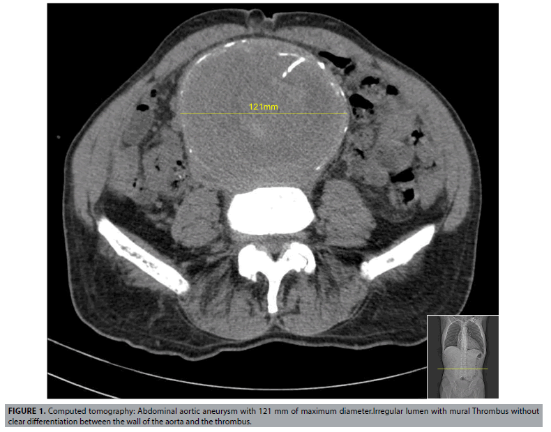
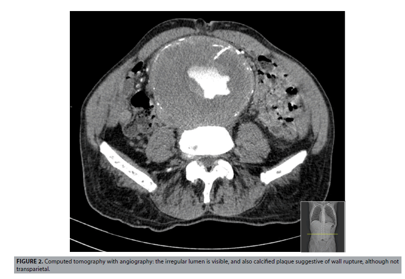
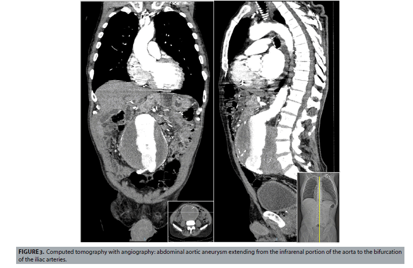
Abdominal Aortic Aneurysms (AAA) frequently develops without causing any symptoms, until they rupture. From that moment on, its prognosis is usually gruesome( FIGURES 1-3)[1].
Caucasian male, 75-years-old, with history of heart failure with severely depressed left ventricle ejection fraction in the setting of dilated cardiomyopathy, hypercholesterolemia and heavy smoking (180 pack-year). Admitted to the emergency department with complaints of fever, dysuria, trembling and 3 episodes of gastric vomit in the previous four hours. On clinical examination he presented with fever (38.5°C), elevated blood pressure (151/83 mmHg) and a pulsatile abdominal mass, located in the central quadrants, with approximately 10 cm of transversal diameter. A thoracic-abdominal-pelvic Computerized Tomography (CT) angiography showed an infra renal abdominal aortic aneurysm with 12 cm of transversal diameter with signs of active intramural hemorrhage and high risk of rupture. All the remaining branches of abdominal aorta were permeable. An Endovascular Repair of the Aneurysm (EVAR) was performed, without any intra-operatory complications. Nevertheless, 24 hours later he presented with acute abdominal pain and a new abdominal-pelvic CT angiography showed an acute occlusion of the Superior Mesenteric Artery (SMA). He was taken to the operatory room but there was no possibility of reversing the extensive intestinal necrosis and he died 24 h hours later. The clinical relevance of this case is mainly based on its atypical presentation. Also, early detection of AAA allows elective repair and improves outcomes, so this too demonstrates the importance of screening for this entity in all men at 65 years of age, in accordance with the most recent guidelines published by the European Society for Vascular Surgery [2]. In addition, the cause of death in this patient was a thromboembolic occlusion of the SMA, a rare post-operative complication in the EVAR era [3].
References
- Kent KC. Abdominal aortic aneurysms. N Engl J Med. 371(22), 2101-2108 (2014).
- Wanhainen A, Verzini F, Van Herzeele I et al . Editor's choice - European Society for Vascular Surgery (ESVS) 2019 clinical practice guidelines on the management of abdominal aorto-iliac artery aneurysms. Eur J Vasc Endovasc Surg. 57(1), 8-93 (2019).
- Daye D, Walker TG. Complications of endovascular aneurysm repair of the thoracic and abdominal aorta: evaluation and management. Cardiovasc Diagn Ther. 8(1), S138-S156 (2018).


