Image - Clinical Practice (2020) Volume 17, Issue 5
An os sustentaculi with an accompanying talocalcaneal synchondrosis: An image articles
- Corresponding Author:
- Elizabeth Swan
Open Access Publishers, 40 Bloomsbury Way
Lower Ground Floor, United Kingdom
Abstract
The sustentaculum bone is a hard prominence at the average surface of the calcaneus that verbalizes with the center calcaneal articular surface of the bone and offers a significant help for the average section of the foot. Its upper articular surface is inward, and its lower surface has a score for the ligament of the flexor hallucis longus. The sustentaculum bone offers connection to the plantar calcaneonavicular (spring) tendon on its foremost edge and the tibiocalcaneal tendon and the average talocalcaneal tendon (part of the deltoid tendon) on its average edge.
Description
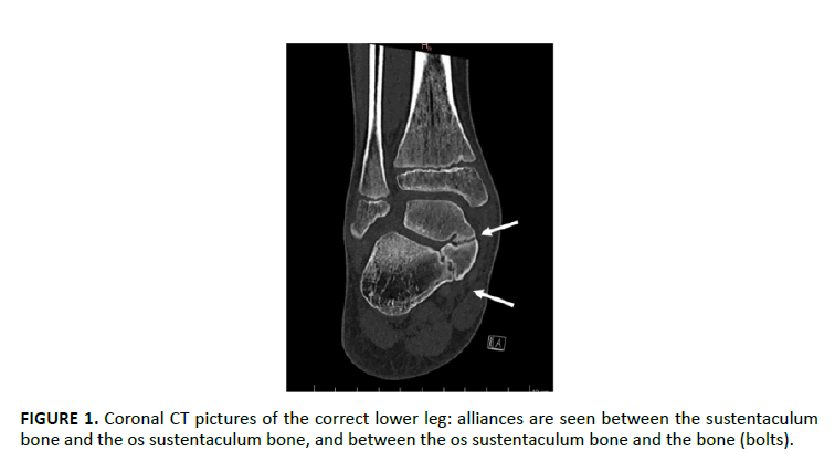
Talocalcaneal Alliance (TCC) is characterized as a joining between the bone and the calcaneus, ordinarily connected with an irregular hypertrophy of the average part of the bone and the sustentaculum bone FIGURE 1. TCC is analyzed in under 1% of everybody. Nonetheless, as the hard variety is frequently asymptomatic the genuine commonness is most likely a lot higher. In the pediatric populace, talocalcaneal alliance is of inherent root and results from a disappointment of mesenchymal division of calcaneus and bone. Generally, this condition gets indicative in the second decade of life. A TCC can introduce itself as a synostosis, as a synchondrosis, or as a syndesmosis. Moreover, it tends to be grouped by its area, as intraarticular (influencing either front, average, or back aspects) or extra articular (normally posteromedial).


