Case Report - Interventional Cardiology (2021)
Combining reverse nano-crush and stentboost technique to optimize outcome for bifurcation stenting
- Corresponding Author:
- Rajeev Chauhan
Department of Cardiology,
Postgraduate Institute of Medical Education and Research,
Chandigarh,
India,
E-mail: rajeevkilroy@yahoo.com
Received date: January 03, 2021; Accepted date: January 28, 2021; Published date: February 04, 2021
Abstract
Though provisional stenting is the strategy of choice for bifurcation lesions; many patients need stenting of both main vessel as well as side branch. In order to ensure optimal stent expansion at ostium of side branch as well as reducing the amount of metal struts inside main vessel, reverse nano-crush is an excellent technique. We report a case of a 75 year old male who presented with ST elevation inferior wall myocardial infarction; whose angiography revealed significant disease in left circumflex and obtuse marginal (major) branch. He underwent percutaneous intervention using reverse nano-crush technique along with utilization of stentboost enhancement to ensure adequate stent expansion. The combination of reverse nano-crush with stentboost enhancement can emerge as the strategy of choice for bifurcation lesions; whenever intracoronary imaging is unavailable or not feasible.
Keywords
Bifurcation stenting • Per-cutaneous Coronary Intervention (PCI)• Reverse Nano-Crush • Ambulatory blood pressure monitoring • Stentboost enhancement
Introduction
Bifurcation stenting is a complex Percutaneous Coronary Intervention (PCI) technique which needs to be performed by experienced operator after meticulous assessment of coronary angiographic images. Various techniques have been described; with each having its inherent advantages and disadvantages. Provisional stenting is considered standard technique for most of the bifurcation lesions unless there is significant ostio-proximal disease in the side branch vessel. Nano crush technique has been recently described for bifurcation lesions with optimal outcomes. Utilization of Stentboost enhancement can provide adequate visualization to assess stent expansion in both main branch as well as side branch. We describe a combining novel technique ‘Reverse Nano-Crush’ with Stentboost enhancement for bifurcation stenting which can provide excellent outcomes.
Case Report
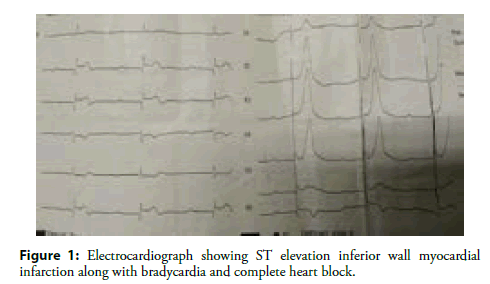
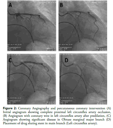
A 75-year-old non-diabetic, non-smoker hypertensive male presented with anginal chest pain of 2 days duration. His pulse was 40 per min with blood pressure of 100/68 mm of Hg. His jugular venous pulse revealed cannon ‘a’ waves. His respiratory and neurological examination was unremarkable. His electrocardiogram ST elevation in leads II, III and aVF along with complete heart block with wide QRS escape complexes (Figure 1). His echocardiography revealed ejection fraction of 45% with regional wall motion abnormality in basal inferior and baso-lateral regions. His troponin levels were raised (15.0 ng/mL). His haematological and biochemical investigations were normal. After informed consent he underwent urgent temporary pacemaker insertion via right femoral vein access. He was started on dual antiplatelet, statin, and tirofiban infusion. He later underwent coronary angiography via right femoral artery access which revealed dominant proximal left circumflex (LCX) artery complete occlusion (Figure 2A). In view of complete heart block it was decided to proceed with revascularization.
Figure 1: Electrocardiograph showing ST elevation inferior wall myocardial infarction along with bradycardia and complete heart block.
Figure 2 : Coronary Angiography and percutaneous coronary intervention (A) Initial angiogram showing complete proximal left circumflex artery occlusion, (B) Angiogram with coronary wire in left circumflex artery after predilation, (C) Angiogram showing significant disease in Obtuse marginal major branch (D) Placement of drug eluting stent in main branch (Left circumflex artery).
Percutaneous coronary intervention was planned after informed consent. Right femoral artery access was taken via 6 Fr femoral sheath and PB‐3.0 6.0 F (Asahi Intec) guiding catheter was used. Coronary wire (‘Runthrough NS’) was successfully crossed across the lesion into distal LCX (Figure 2B). The lesion was predilated with semi-compliant balloons (Ikazuchi 1.0 × 12 mm and Pantera Pro 2.0 × 20 mm). The Obtuse Marginal Major (OMM) branch was of significant size, hence another coronary wire was placed in OMM. Thereafter drug eluting stent ‘Abluminus’ (3.0 × 28 mm) was placed in Main Branch (MB) i.e. LCX across the lesion. However, angiography revealed significant disease in ostioproximal OMM (Figure 2C). Hence it was planned to utilize novel technique ‘Reverse Nano-Crush technique’ to place stent across Side Branch (SB).
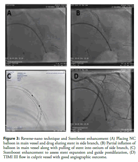
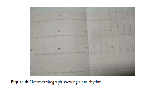
NC Emerge balloon (2.75 × 15 mm) was placed in MB inside the stent and drug eluting stent (Orsiro 2.75 × 15 mm) was placed in SB from ostium (Figure 3A). The balloon in MB was partially inflated and stent in SB was pulled across ostium into MB; in order to ensure that proximal part of stent covers ostium without significant amount of struts in MB (Figure 3B). The stent was deployed at nominal pressure. Both stent balloon in SB and NC balloon in MB were deflated simultaneously. Now stent balloon was carefully pulled across ostium and re-inflated to open the struts at ostium. Thereafter standard kissing balloon inflation was carried out followed by proximal optimization of MB stent. Stentboost enhancement was utilized to assess stent expansion and accordingly post dilation of the stents was carried out (Figure 3C). The final angiographic result showed excellent stent expansion without significant struts of SB stent in MB along with TIMI III flow (Figure 3D). Patient recovered gradually and over his complete heart block also resolved and TPI was removed. Patient was continued on dual antiplatelet, statin and supportive care. He remains asymptomatic at 1 month follow up with sinus rhythm (Figure 4).
Figure 3: Reverse-nano technique and Stentboost enhancement (A) Placing NC balloon in main vessel and drug eluting stent in side branch, (B) Partial inflation of balloon in main vessel along with pulling of stent into ostium of side branch, (C) Stentboost enhancement to assess stent expansion and guide postdilatation, (D) TIMI III flow in culprit vessel with good angiographic outcome.
Results and Discussion
The coronary bifurcations are vulnerable for atheroma development due to turbulent flow pattern and endothelial shear stress [1]. The bifurcation stenting is associated with lower procedural success rates and increased long term adverse cardiac events [2]. The stenting of main vessel alone; if feasible has been associated with better clinical outcomes like stent thrombosis as compared to stenting across both MB and SB [3]. Thus; bifurcation lesion are anatomically complex and require high level of technical expertise to achieve optimal clinical outcomes. Provisional stenting strategy is considered superior to upfront two stent strategy as provisional stenting offers better long term mortality outcomes [4]. After stenting of MB; SB should be treated if antegrade flow is impaired (TIMI flow <3), when severe ostial SB narrowing is present and fractional flow reserve of the SB is <0.80 [5]. If SB needs stenting options are: T/TAP stenting, Culotte stenting, DK-Crush and Mini-Crush technique. All these techniques have inherent advantages and disadvantages based on coronary anatomy complexity and lesion characteristics. We used a Nano-Crush technique with slight modification; which is called as ‘Reverse Nano-Crush’ technique [6].
On artificial coronary flow models using computational fluid dynamics, among various bifurcation stenting strategies, Nano-crush and modified T techniques had achieved the most physiologic profile [7]. In a study on 205 patients who underwent Nano-Crush bifurcation stenting, bench study and computational fluid dynamics evaluation suggested a complete coverage of the SB ostium with a very high strut-free area at the SB [8]. In a retrospective study on 65 patients by Rigatelli G et al found that nano-crush technique was associated with less use of contrast, less procedural time and less X-ray exposure compared to the culotte technique for the treatment of unprotected left main bifurcation lesions [9]. In fact, in a study on 752 patients with STEMI and cardiogenic shock who underwent left main bifurcation stenting, it was observed that Nano-crush had better 3 year cardiovascular mortality outcomes as compared to T-stenting [10]. The combination of newer stents with ultra-thin struts and newer bifurcation stenting strategies like nano-crush can provide good short as well as long term clinical outcomes in bifurcation stenting especially involving left main vessel [11].
Intracoronary imaging is currently being utilized in various coronary interventions; however, its use is limited by significantly increased cost, procedural time, and the need for additional training of staff. Stentboost is a simple, fast, and cost-effective tool to enhance stent visualization by enhancing the X-ray focus of the region where stent is placed [12]. Stentboost enhancement has shown better correlations for stent expansion measured by intravascular ultrasound imaging when compared with quantitative coronary angiography [13]. Utilizing stentboost can reduce cost as well as time of procedure while simultaneously ensuring adequate stent expansion. We utilized stentboost enhancement in bifurcation stenting to guide postdilation of the stents. The Stentboost has been utilized in bifurcation stenting with good outcomes and also in dedicated bifurcation stents. However, in our knowledge this was first case of utilizing Reverse Nano-Crush technique along with stentboost enhancement to ensure optimal outcomes in Bifurcation stenting. In order to assess wider applicability and outcome of this technique; a study with larger sample size needs to be carried out.
Conclusion
Nano-crush technique is an excellent option for bifurcation stenting. Reverse Nano-crush can appear as first choice technique in provisional stenting strategy whenever SB needs to be stented as this technique leads to optimal SB ostium coverage by stents as well as minimal struts inside the MB. Combining stentboost enhancement with reverse nano-crush technique can be best strategy for most of bifurcation lesions.
References
- Giannoglou GD, Antoniadis AP, Koskinas KC, et al. Flow and atherosclerosis in coronary bifurcations. EuroIntervention. 6(Supplement J); J16-J23 (2010).
- Steigen TK, Maeng M, Wiseth R, et al. Randomized study on simple versus complex stenting of coronary artery bifurcation lesions: The Nordic bifurcation study. Circulation 114(18): 1955-61 (2006).
- Katritsis DG, Siontis GC, Ioannidis JP. Double versus single stenting for coronary bifurcation lesions: a meta-analysis. Circ Cardiovasc Interv. 2(5): 409-15 (2009).
- Nairooz R, Saad M, Elgendy IY, et al. Long-term outcomes of provisional stenting compared with a two-stent strategy for bifurcation lesions: A meta-analysis of randomised trials. Heart. 103(18): 1427-1434 (2017).
- Sawaya FJ, Lefèvre T, Chevalier B, et al. Contemporary approach to coronary bifurcation lesion treatment. JACC Cardiovasc Interv. 9(18): 1861-78 (2016).
- Rigatelli G, Dell'Avvocata F, Zuin M, et al. Complex coronary bifurcation revascularization by means of very minimal crushing and ultrathin biodegradable polymer DES: Feasibility and 1-year outcomes of the "Nano-crush" technique. Cardiovasc Revasc Med. 18(1): 22-27 (2017).
- Rigatelli G, Zuin M, Dell'Avvocata F, et al. Complex coronary bifurcation treatment by a novel stenting technique: Bench test, fluid dynamic study and clinical outcomes. Catheter Cardiovasc Interv. 92(5) 907-914 (2018).
- Rigatelli G, Zuin M, Vassilev D, et al. Culotte versus the novel nano-crush technique for unprotected complex bifurcation left main stenting: Difference in procedural time, contrast volume and X-ray exposure and 3-years outcomes. Int J Cardiovasc Imaging. 35(2): 207-214 (2019).
- Rigatelli G, Zuin M, Dinh H, et al. Long-Term outcomes of left main bifurcation double stenting in patients with STEMI and cardiogenic shock. Cardiovasc Revasc Med. 20(8): 663-668 (2019).
- Koolen JJ, van het Veer M, Hanekamp CE. Stentboost image enhancement: First clinical experience. Medicamundi 49(2): 4-8 (2005).
- Laimoud M, Nassar Y, Omar W, et al. Stent boost enhancement compared to intravascular ultrasound in the evaluation of stent expansion in elective percutaneous coronary interventions. Egypt Heart J. 70(1): 21-26 (2018).
- Silva JD, Carrillo X, Salvatella N, et al. The utility of stent enhancement to guide percutaneous coronary intervention for bifurcation lesions. EuroIntervention. 9(8): 968-74 (2013).
- Fysal Z, Hyde T, Barnes E, et al. Evaluating stent optimisation technique (StentBoost®) in a dedicated bifurcation stent (the Tryton™). Cardiovasc Revasc Med. 15(2): 92-6 (2014).
- Roik M, Wretowski D, Wolny R, et al. StentBoost imaging for the assessment of optimal stent deployment and coverage of side branch ostium in coronary bifurcation intervention. Int J Cardiol. 172(3): e458-60 (2014).


