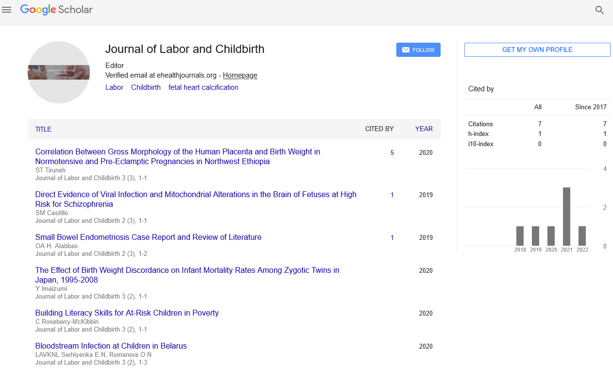
Mini Review - Journal of Labor and Childbirth (2022) Volume 5, Issue 5
Fetal Cardiac Calcifications: Report of Four Prenatally Diagnosed Cases and Review of the Literature
Moatasem Bellah Al Farrah*
Department of Gynecologist, Charles University, Germany
Received: 01-Aug -2022, Manuscript No. JLCB-22-72826; Editor assigned: 03-Aug-2022, PreQC No. JLCB-22- 72826 (PQ); Reviewed: 17- Aug -2022, QC No. JLCB-22-72826; Revised: 22- Aug -2022, Manuscript No. JLCB-22- 72826 (R); Published: 31- Aug -2022, DOI: 10.37532/jlcb.2022.5(5).77-81
Abstract
Fetal cardiac calcifications are defined as diffuse hyperechogenicities in the different layers
of the heart. This is an uncommon fetal ultrasound finding associated with significant
myocardial dysfunction. We report four cases with massive fetal myocardial calcifications
detected on prenatal ultrasound at 18-22 weeks’ gestation and associated, in all cases, with
significant cardiac dysfunction. Detailed fetal echocardiographic evaluation, chromosome
analysis, and an extensive search for intrauterine infection as a cause of these abnormalities,
were carried out on all cases.
Introduction
A thorough autopsy was performed on all deceased fetuses and postnatal investigation of the sole survivor was performed. Two of our patients chose to interrupt their pregnancies, one fetus suffered intrauterine demise, and one child was born alive. In all of our cases the karyotypes were normal, and no specific infectious etiology or maternal autoantibody was noted. Histopathology findings in the non-survivors included myo- and epicardial calcification maximal at the base of the heart [1]. The living child has findings suggestive of an intrauterine infection, although no infectious entity was identified. Long-term follow-up showed sensorineural hearing loss and severe developmental delay.
One-hundred and fifty-one fetuses with calcifications and 302 matched controls were selected from the archives of the Department of Pathology, Karolinska University Hospital. Chromosome analysis by karyotyping or quantitative fluorescence-polymerase chain reaction was performed. Autopsy and placenta reports were scrutinized for presence of malformations and signs of infection [2].
Calcifications were mainly located in the liver, but also in heart, bowel, and other tissues. Fetuses with calcifications showed a significantly higher proportion of chromosomal abnormalities than controls; 50% vs. 20% (p<0.001). The most frequent aberrations among cases included trisomy 21 (33%), trisomy 18 (22%), and monosomy X (18%). A similar distribution was seen among controls. When comparing cases and controls with chromosomal abnormalities, the cases had a significantly higher prevalence of malformations (95% vs. 77%, p=0.004). Analyzed the other way around, cases with malformations had a significantly higher proportion of chromosomal abnormalities compared with controls, (66% vs. 31%, p<0.001) [3].
The presence of calcifications in fetal tissues is occasionally recognized both at autopsy and on ultrasound imaging, but their biological importance remains poorly understood. At autopsy, calcifications are identified on histological sections or even macroscopically, if sufficiently large. On ultrasound they are recognized as hyperechogenic sites, which echogenicity resembles that of the surrounding bone [4].
Previous studies have mainly focused on liver calcifications, which have been reported in 2.2% to 4.2% of cases in autopsy studies and with an estimated incidence ranging from 1:260 to 1:1750 in ultrasound screening. When identified by ultrasound, cases with calcification as the only aberrant finding usually have a good outcome, i.e. the birth of a healthy child. However, when identified together with other abnormalities, the prognosis is poor. Studies have suggested association of calcifications with infection, circulatory compromise and chromosomal abnormalities [5].
Fetal liver calcifications have been identified in number cases of trisomy 18 as well as in cases of other aneuploidies. Additionally, a high incidence of various chromosomal abnormalities has been identified in fetuses with calcifications located in the heart. Taken together, the association between fetal tissue calcifications and chromosomal abnormalities has been indicated in previous studies. Here we explore this association by a matched case-control study [6].
The study included 151 fetuses with calcifications and 302 matched controls. The cases were retrospectively identified from the archives of the Center for Perinatal Pathology at the Department of Pathology, Karolinska University Hospital, corresponding to all cases with registered fetal calcifications from January 1, 2003 to December 31, 2012. All histological sections were re- examined by two perinatal pathologists to verify the presence of calcifications [7]. All sections were originally stained with Hematoxylin and Eosin, according to standard procedure. In several dubious cases, special staining (von Kossa) was applied to verify the presence of calcifications. Fetuses from the same archives with the closest analysis date before and after each case, were selected as controls and matched for gestational age (GA) and type of death (spontaneous or missed abortion, stillbirth, induced termination of pregnancy). Missed abortion was defined as fetal death in utero (up to gestational week 21+6) that had not been followed by immediate expulsion. Stillbirth was defined as fetal death occurring later than gestational week 22+0. Autopsy and placenta reports for all study subjects were scrutinized with focus on the presence of malformations and signs of infection. Malformation was defined as major structural anomaly in the fetus; for example, minor dysmorphism, isolated abnormal lung lobation, simian crease or simple ectopia of an organ or a tissue was not included. Signs of infection, irrespective of gestational age, were sought for in the placenta (acute chorioamnionitis, vasculitis or funisitis, representing bacterial infection, or chronic villitis, representing viral infection) or the fetus (most often bronchopneumonia). In some cases of stillbirth the infection was corroborated by positive bacterial culture. Viral infection (most notably cytomegalovirus) was in some cases documented by immunohistochemistry or positive viral serology [8].
Chromosome analysis by conventional karyotyping or quantitative fluorescence- polymerase chain reaction (QF-PCR) had previously been performed on 290 of the 453 fetuses included in the study, according to analysis results from the archives of the Clinical Genetics Unit, Karolinska University Hospital [9]. For the remaining fetuses, tissue samples were collected from the biobank of the Department of Perinatal Pathology, Karolina University Hospital, for complementary analysis by QF-PCR. For cases analysed by karyotyping, at least 11 metaphase nuclei per sample were analysed with conventional Q-banding, using standard cytogenetic procedures. In cases where cell culturing was unsuccessful, and for the samples collected retrospectively in the biobank, DNA was extracted from amniotes, chorionic villi, or fetal tissue using the Instance Matrix protocol (Bio-Rad), and analysed using a QF-PCR panel for detection of aneuploidies involving chromosomes 13, 18, 21, X and Y as previously described [10].
McNamara’s test for matched case-control studies was used to determine statistically significant differences between proportions of chromosomal abnormalities and malformations in cases and controls, as well as in subgroups (gestational age intervals, different types of death, and different tissue locations of calcifications). The significance level of all analyses was set to 0.05. However, as each case was matched with two controls, a Bonferroni correction of the significance level was made; hence the significance level was 0.025 in the McNamara calculation. A chi-squared test was used to assess statistically significant differences in distribution of identified chromosomal abnormalities in cases and controls, as well as to detect significant differences in the amount of malformations and signs of infection between the groups. All calculations were performed using IBM Statistical Package for the Social Sciences (SPSS) version 21 [11].
The overall proportion of fetuses with calcifications in the archives was 5.3%. The proportion showed a steady increase over the years of analysis, from 3.1% in 2003, to 8.2% in 2012. The highest proportion of calcifications was seen among fetuses in gestational week 13–15, where it exceeded 10%. Calcifications were mainly located in the liver (57%), but also in heart (13%), bowel (6%) and other tissues. Calcifications in multiple tissues were identified in 22% of the cases. Fetuses with calcifications showed a significantly higher proportion of chromosomal abnormalities compared with controls, 50% vs. 20% (p<0.001) [12]. The proportion of chromosomal abnormalities in all subgroups is summarized in. For subgroups based on gestational age intervals, the highest proportion of chromosomal abnormalities was seen in cases of gestational age (GA) <14 (71%) and 23–28 (75%), although the number of cases was too low to reach statistical significance in the latter group. The lowest proportion of chromosomal abnormalities was identified in fetuses of GA >29, and no significant difference was detected between cases and controls in this subgroup (17% vs. 13% in cases and controls, respectively) [13]. For subgroups based on type of death, the highest proportion of chromosomal abnormalities was detected among cases after induced termination, where both cases and controls had a higher proportion than the average (63% and 34%, respectively). No significant difference was detected between cases and controls in the stillbirth group, and no chromosomal abnormalities at all were detected in the spontaneously aborted fetuses. However, the number of cases was low in both of these subgroups. The tissue location of calcifications did not influence the proportion of chromosomal abnormalities identified [14].
We describe the first matched case-control study on fetal tissue calcifications, in which we show an association between calcifications and chromosomal abnormalities; 50% vs. 20% in cases and controls, respectively. When creating subgroups based on type of death, the highest proportion of chromosomal abnormalities in both cases and controls was identified in terminated pregnancies (63% and 34%, respectively). This was expected as the main reason for pregnancy termination followed by autopsy is a fetal chromosomal abnormality. The lowest proportion, 31% in cases and 9% in controls, was found in the stillbirth group, except from the subgroup of spontaneously aborted fetuses, where no aberrations were identified. However, as the spontaneous abortion group included only six cases, no conclusions can be drawn [15]. The likely explanation for the low number of chromosomal abnormalities in the stillbirth group is that fetal chromosomal abnormalities is not as frequent after gestational week 22 compared with earlier in pregnancy, as the vast majority of fetuses with chromosomal abnormalities are spontaneously aborted or detected and terminated earlier in pregnancy . This is also reflected in the subgroups based on gestational age; the proportion of chromosomal abnormalities decreases from 71% of cases of GA <14 to 17% in cases of GA >29 (with the exception of fetuses of GA 23–28). The distribution of identified chromosomal abnormalities did not differ significantly between cases and controls. We noted a tendency that trisomy 13 and 18 was more frequent in cases than in controls, but a larger cohort would be required to establish a true difference in distribution [16].
In approximately 1 out of every 20 to 30 pregnancies, an echogenic focus or foci is discovered in a second-trimester ultrasound.1 These bright spots seen in the heart are called echogenic intracardiac foci (multiple) or an echogenic intracardiac focus (singular), which is often shortened to EIF, a cardiac echogenic focus, or echogenic focus [17].
This condition is considered a normal variation and generally doesn’t affect the baby’s heart or its functioning. On ultrasound, there might be one or more bright spots found, usually in the ventricles, which pump blood. It is not a heart defect and for the majority of instances in which this occurs, it poses no risk to the fetus [18].
While the EIF might disappear during the third trimester, many times it is still present on later ultrasounds. Follow-up imaging studies aren’t typically recommended unless other abnormalities are found on the ultrasound and/or the pregnancy is at higher risk for chromosomal anomalies. In fact, echogenic focus is found in 3% to 5% of normal pregnancies [19].
A limitation of this study is that two different methods were used for chromosome analysis. Among the cases, 48% were analyzed by conventional karyotyping and 52% by QF-PCR. The corresponding numbers among controls were 38% and 62%, respectively. A drawback of QF-PCR is that it only gives information about a limited number of chromosomes, and that no structural aberrations can be detected. In fetuses analyzed by karyotyping, the proportion of detected aberrations was increased by 5.6% and 5.2% in cases and controls, respectively, compared with if the same fetuses would have been analyzed by QF-PCR only [20]. If the complete study cohort had been analyzed by karyotyping, the proportion of detected aberrations could potentially have increased from 50% to 54% among cases and from 20% to 24% among controls, assuming that the 5.6% and 5.2% increase rate would hold true in fetuses analyzed by QF-PCR in our study. Although karyotype analysis of all fetuses most probably would have identified an additional number of chromosomal abnormalities, QF-PCR still shows its great value as a complementary analysis when karyotype is unsuccessful or not suitable, as it has the potential to identify the vast majority of cases with chromosomal abnormalities [21,22].
Discussion
In summary, we have shown that fetal tissue calcifications are highly associated with chromosomal abnormalities in combination with congenital malformations. Identification of a calcification together with a malformation at autopsy more than doubles the probability of detecting a chromosomal abnormality, compared with identification of a malformation only. We propose that identification of a fetal tissue calcification at autopsy, and potentially also at ultrasound examination, should infer special attention towards co-existence of malformations, as this would be a strong indicator for a chromosomal abnormality [23].
In the majority of patients, the echogenic intracardiac focus is representative of a calcification or a microscopic fibrosis inside the papillary muscle. The presence of the bright spot on the ultrasound imaging may also be another condition altogether. For instance, fetal cardiac tumours like rhabdomyomas may become calcified and show up as an echogenic intracardiac focus. This is the reason for referring the fetus for careful echocardiography. Another condition which can mimic the appearance of an echogenic intracardiac focus is endocardial fibroelastosis, which is a rare heart disease found in babies. Here, the endocardiaum of the fetal heart becomes calcified or is affected by fibrosis. This causes the muscular lining of the heart c [24].
It is important to remember that the presence of an echogenic intracardiac focus is not unusual. Routine fetal ultrasounds may show this condition, which may be a reason to refer the pregnant woman for fetal echocardiography. This second-level test is performed to check for the risk of congenital heart defects in the infant.
Often the pregnant woman will be asked to undergo an amniocentesis, should the health care professional suspect a genetic or other problem with the fetus.
The echogenic intracardiac focus is usually caught on an ultrasound examination in the first trimester ( about 14 weeks of pregnancy). In some cases, the condition disappears by the time the pregnant woman comes in for her next ultrasound in the second trimester.
Conclusion
However, in the majority of cases the condition persists into the third trimester, even though its size is considerably reduced by this time. It typically disappears towards the latter part of the third trimester. In normal pregnancies the presence of an echogenic intracardiac focus is viewed as a benign variant. However, in high risk pregnancies it may be seen as a soft marker for aneuploidy anomalies such as Down syndrome and trisomy 13. The presence of multiple echogenic intracardiac foci may be seen when there is an increased risk of congenital heart disease.
Acknowledgement
None
Conflict of Interest
There is no conflict of Interest.
References
- Acherman RJ, Evans WN, Luna CF et al. Prenatal detection of congenital heart disease in southern Nevada: the need for universal fetal cardiac evaluation. J Med Ultrasound. 26, 1715-1719 (2007).
- Bahtiyar MO, Copel JA. Improving detection of fetal cardiac anomalies: a fetal echocardiogram for every fetus? J Med Ultrasound. 26, 1639-1641 (2007).
- Carvalho JS. Early prenatal diagnosis of major congenital heart defects. Curr Opin Obstet Gynecol. 13, 155-159 (2001).
- Davey BT, Seubert DE, Phoon CK et al. Indications for fetal echocardiography high referral, low yield? Obstet Gynecol Surv. 64,405-415 (2009).
- Lee W, Allan L, Carvalho JS et al. ISUOG consensus statement: what constitutes a fetal echocardiogram? Ultrasound Obstet Gynecol. 32,239-242 (2008).
- Hoffman JI, Kaplan S. The incidence of congenital heart disease. J Am Coll Cardiol. 39, 1890-1900 (2002).
- Vos T, Allen C, Arora M et al. Global, regional, and national incidence, prevalence, and years lived with disability for 310 diseases and injuries, 1990-2015: a systematic analysis for the Global Burden of Disease Study 2015. Lancet. 388, 1545-1602 (2016).
- Wang H, Naghavi M, Allen C et al. Global, regional, and national life expectancy, all-cause mortality, and cause-specific mortality for 249 causes of death, 1980-2015: a systematic analysis for the Global Burden of Disease Study 2015. Lancet. 388, 1459-1544 (2016).
- Blue GM, Kirk EP, Giannoulatou E et al. Advances in the Genetics of Congenital Heart Disease: A Clinician's Guide. J Am Coll Cardiol. 69, 859-870 (2017).
- Costain G, Silversides CK, Bassett AS et al. The importance of copy number variation in congenital heart disease. NPJ Genom Med. 1, 16031 (2016).
- Bouma BJ, Mulder BJ. Changing Landscape of Congenital Heart Disease. Circ Res.120, 908-922 (2017).
- Zaidi S, Brueckner M. Genetics and Genomics of Congenital Heart Disease. Circ Res. 120, 923-940 (2017).
- Razmara E, Garshasbi M. Whole-exome sequencing identifies R1279X of MYH6 gene to be associated with congenital heart disease. BMC Cardiovasc Disord. 18, 137 (2018).
- Cantù C, Felker A, Zimmerli D et al. Pygo genes cause congenital heart defects by tissue-specific perturbation of Wnt/β-catenin signaling. Genes Dev. 32, 1443-1458 (2018).
- Canobbio MM, Warnes CA, Aboulhosn J et al. Management of Pregnancy in Patients With Complex Congenital Heart Disease: A Scientific Statement for Healthcare Professionals From the American Heart Association. Circulation. 135, e50-e87 (2017).
- Lozano R, Naghavi M, Foreman K et al. Global and regional mortality from 235 causes of death for 20 age groups in 1990 and 2010: a systematic analysis for the Global Burden of Disease Study 2010. Lancet. 380, 2095-128 (2012).
- Spitzer A. Uber den Bauplan des normalen und missbildeten Herzens: Versuch einer phylogenetischen Theorie. Virchows Arch Pathol Anat. 243, 81-272 (1923).
- Rasmussen SA, Galuska DA. Prepregnancy obesity and birth defects: what's next? Am J Clin Nutr. 91, 1539-1540 (2010).
- Mills JL, Troendle J, Conley MR et al. Maternal obesity and congenital heart defects: a population-based study. Am J Clin Nutr. 91, 1543-1549 (2010).
- Zhang S, Wang L, Yang T et al. Parental alcohol consumption and the risk of congenital heart diseases in offspring: An updated systematic review and meta-analysis. Eur J Prev Cardiol. 27, 410-421 (2020).
- Tidyman WE, Rauen KA. The RASopathies: developmental syndromes of Ras/MAPK pathway dysregulation. Curr Opin Genet. 19, 230-236 (2009).
- Niessen K, Karsan A. Notch signaling in cardiac development. Circ Res. 102, 1169-1181 (2008).
- Srivastava D. Making or breaking the heart: from lineage determination to morphogenesis. Cell. 126, 1037-1048 (2006).
- Vos T, Barber RM, Bell et al. Global, regional, and national incidence, prevalence, and years lived with disability for 301 acute and chronic diseases and injuries in 188 countries, 1990-2013: a systematic analysis for the Global Burden of Disease Study 2013. Lancet. 386 (9995): 743-800 (2015).
Google Scholar, Crossref, Indexed at
Google Scholar, Crossref, Indexed at
Google Scholar, Crossref, Indexed at
Google Scholar, Crossref, Indexed at
Google Scholar, Crossref, Indexed at
Google Scholar, Crossref, Indexed at
Google Scholar, Crossref, Indexed at
Google Scholar, Crossref, Indexed at
Google Scholar, Crossref, Indexed at
Google Scholar, Crossref, Indexed at
Google Scholar, Crossref, Indexed at
Google Scholar, Crossref, Indexed at
Google Scholar, Crossref, Indexed at
Google Scholar, Crossref, Indexed at
Google Scholar, Crossref, Indexed at
Google Scholar, Crossref, Indexed at
Google Scholar, Crossref, Indexed at
Google Scholar, Crossref, Indexed at
Google Scholar, Crossref, Indexed at

