Image Article - Imaging in Medicine (2022) Volume 14, Issue 6
Folding Fan Sign of Closed-Loop Obstruction
Koki Kawanishi1*, Yoshifumi Ikeda1, Masayuki Kitano2
1Department of Gastroenterology, Nate Hospital, 294-1 Nate Ichiba, Kinokawa City, Wakayama 649-6631, Japan
2Second Department of Internal Medicine, Wakayama Medical University, 811 Kimiidera, Wakayama City, Wakayama 641-0012, Japan
- *Corresponding Author:
- Koki Kawanishi
Department of Gastroenterology, Nate Hospital, 294-1 Nate Ichiba, Kinokawa City, Wakayama 649-6631, Japan
E-mail: amurosyaa218@yahoo.co.jp
Abstract
We coined the new term folding fan sign on abdominal contrast-enhanced computed tomography for strangulated ileus caused by closed-loop obstruction. It can immediately indicate the necessity for surgical intervention.
Keywords
Beak sign • closed loop • ileus
Case Description
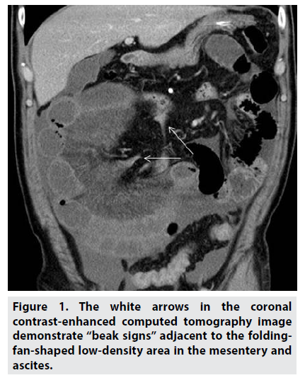
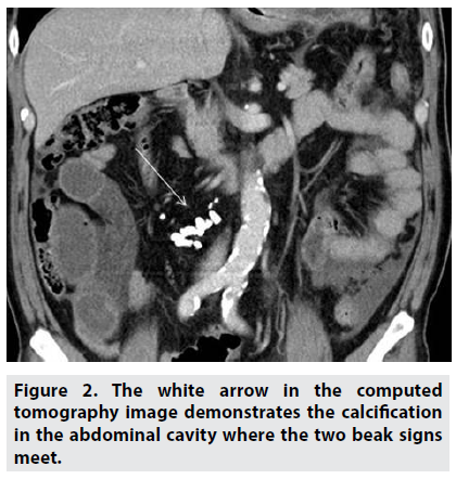
A 73-year-old man was admitted to our hospital because of severe abdominal pain and bilious vomiting. Contrast-enhanced computed tomography (CECT) revealed a dilated small bowel loop. The coronal CECT image demonstrates the so-called beak signs (Figure 1, arrow) adjacent to the folding-fan-shaped lowdensity area in the mesentery and ascites. An emergency surgery was performed. The ascites was hemorrhagic, and an approximately 120-cm segment of the jejunum and ileum was herniated through a slit-like defect in the omentum. The strangulated small bowel loop was removed with an adhesive band. The folding-fan-shaped lowdensity area on CECT, which we named “folding fan sign,” turned out to be a severely congested mesentery with hemorrhagic change. Computed tomography image demonstrated a calcification in the abdominal cavity at the area where the two beak signs met (Figure 2, arrow). This calcification and adhesive band might have developed owing to inflammation from tuberculosis, as the patient had a history of lung tuberculosis.
Figure 1: The white arrows in the coronal contrast-enhanced computed tomography image demonstrate “beak signs” adjacent to the foldingfan- shaped low-density area in the mesentery and ascites.
In conclusion, the folding fan sign on CECT in patients with ileus can provide physicians with information of the immediate necessity for surgical intervention because it can serve as a marker of strangulated ileus from closed-loop obstruction.
Author contributions
Koki Kawanishi: collected clinical data, conceptualized the study, and proofread the final manuscript for submission. Yohifumi Ikeda and Masayuki Kitano: critically revised the manuscript.
Conflict of Interest
The authors report no conflict of interest.
Funding Information
There is no funding related to this work.
Data Availability Statement
The data that support the findings of this study are available from the corresponding author upon reasonable request.
Informed Consent
Informed patient consent was obtained for publication of the case details.


