Image - Clinical Practice (2021) Volume 18, Issue 3
Giant infected hydronephrosis: A kidney bag filled with 6300 cc pus
- Corresponding Author:
- Ankur Mittal
All India Institute of Medical Sciences, India
E-mail: drmittal.ankur@gmail.com
Abstract
Giant hydronephrosis has been variably defined in literature as that one that contains more than 1 liter of fluid or comprising >1.6% of the body weight. It is also described as that occupying a hemiabdomen, crossing the midline and occupying more than five vertebral heights. Congenital uretero-pelvic junction obstruction is the most common cause of giant hydronephrosis in children and young adults. Other important causes include vesico-ureteric junction obstruction, ureteral atresia and primary obstructed megaureter. We herein report a rare case of giant hydronephrosis secondary to pelvi-ureteric junction obstruction presenting as an acute abdomen in a young female.
Description
Giant hydronephrosis has been variably defined in literature as that one that contains more than 1 liter of fluid or comprising >1.6% of the body weight [1]. It is also described as that occupying a hemiabdomen, crossing the midline and occupying more than five vertebral heights [2]. Congenital uretero-pelvic junction obstruction is the most common cause of giant hydronephrosis in children and young adults. Other important causes include vesicoureteric junction obstruction, ureteral atresia and primary obstructed megaureter [3]. We herein report a rare case of giant hydronephrosis secondary to pelvi-ureteric junction obstruction presenting as an acute abdomen in a young female.
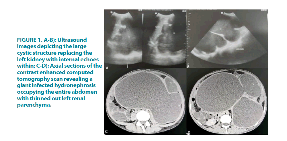
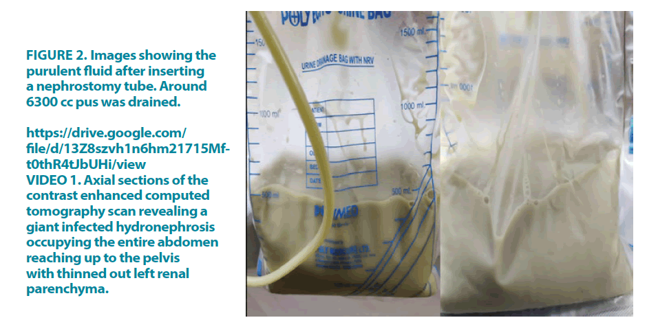
A 22-year-old female presented with complaints of left flank pain, abdominal distention, abdominal discomfort, nausea, vomiting and fever with chills for the past two months. Her general physical examination was unremarkable and her vitals were within normal limits. On examination, abdomen was found to be distended and tense with diffuse tenderness present all over the abdomen with guarding present. A large lump 20 cm × 15 cm lump was palpable in the left side of abdomen extending towards the right iliac fossa and pelvis. An erect chest x-ray was normal with no air under diaphragm. Her hemoglobin was 8.9 g/dl, leucocytes 7600/mm3, serum creatinine was 0.68 g/dl. An ultrasound revealed a large hypoechoic lesion arising from the left kidney (FIGURE 1). A contrast enhanced computed tomography scan was done which showed a giant infected hydro nephrosis of the left kidney measuring (35 × 24 × 14) cm occupying almost the entire abdomen and reaching up to the pelvis secondary to a pelviureteric junction obstruction (FIGURE 1 and VIDEO 1). The parenchyma of the kidney was grossly thinned out (4 mm) and minimal post contrast enhancement. It caused displacement of spleen, stomach, pancreas and hemi diaphragm. No contrast excretion was noted from the left kidney. A 12 Fr percutaneous nephrostomy was placed under antibiotic cover to decompress the kidney and relieve the patient of her symptoms. Frank purulent fluid started draining with over 5800 cc stat output. It continued to drain for the next several hours and completely dried up in 36 hours with a total output of 6300 cc (FIGURE 2). The patient was conservatively managed with intravenous fluids and broadspectrum antibiotics. On follow up examination, the abdomen was soft and there was no tenderness. Microbiological examination of the pus revealed growth of E. coli species. Fungal and tubercular work up was negative. The patient is planned for a left simple nephrectomy.
Figure 1 A-B): Ultrasound images depicting the large cystic structure replacing the left kidney with internal echoes within; C-D): Axial sections of the contrast enhanced computed tomography scan revealing a giant infected hydronephrosis occupying the entire abdomen with thinned out left renal parenchyma.
VIDEO 1. Axial sections of the contrast enhanced computed tomography scan revealing a giant infected hydronephrosis occupying the entire abdomen reaching up to the pelvis with thinned out left renal parenchyma.
Several cases of giant hydronephrosis have been reported, although rarely has a case been reported with over six liters of pus in a kidney presenting as acute abdomen. Giant hydronephrosis usually presents as abdominal discomfort and slowly increasing abdominal distention with episodes of pain. Ultrasound and CT scan usually clinch the diagnosis. The treatment depends on the functional status of the kidney. While reconstructive procedures are performed for functional kidneys, nephrectomy remains the treatment of choice for nonfunctional kidneys. Percutaneous nephrostomy is usually done in such large hydronephrotic kidneys as a temporizing measure [4].
Learning Points/Take Home Messages
• Although rare, UPJO may rarely be silent over many years to finally present as an acute abdomen due to an unusually large infected hydronephrosis occupying almost the entire abdomen
• Percutaneous nephrostomy should be used as a temporizing measure in such situations to prevent intraperitoneal rupture and consequent sepsis
• Giant infected hydronephrosis with nonfunctional thinned out parenchyma usually warrants a nephrectomy once the clinical condition of the patient improves
References
- Wang QF, Zeng G, Zhong L, et al. Giant hydronephrosis due to ureteropelvic junction obstruction: a rare case report, and a review of the literature. Mol Clin Oncol. 5: 19-22 (2016).
- Kaura KS, Kumar M, Sokhal AK, et al. Giant hydronephrosis: Still a reality!. Turk J Urol. 43: 337-344 (2017).
- Shah SA, Ranka P, Dodiya S, et al. Giant hydronephrosis: What is the ideal treatment? Indian J Urol. 20: 118-122 (2004).
- Joseph M, Darlington D. Acute presentation of giant hydronephrosis in an adult. Cureus. 12: e8702 (2020).


