Case Report - Imaging in Medicine (2022) Volume 14, Issue 9
GIANT PRIMARY RETROPERITONEAL DEDIFFERENTIATED LIPOSARCOMA OBSERVED IN BONE SCINTIGRAPHY WITH 99mTc-MDP. CASE REPORT
Nathalie Hernandez-Hidalgo*,Diana C Quijano G, Nikolai Strusberg, Humberto Varela
Unidad de Medicina Nuclear Clínica Palermo, Clínicas Colsanitas, Bogotá, Colombia, Fundacion Universitaria Sanitas, Bogota, Colombia
Unidad de Medicina Nuclear Clínica Palermo, Clínicas Colsanitas, Bogotá, Colombia, Fundacion Universitaria Sanitas, Bogota, Colombia
E-mail: natha8830@gmail.com
Received: 12-Apr-2021, Manuscript No. FMIM-21-29467; Editor assigned: 14-Apr-2022, PreQC No. FMIM-21-29467 (PQ); Reviewed: 01-Feb-2021, QC No. FMIM-22-29467; Revised: 22-Feb-2021, Manuscript No. FMIM-21-29467 (R); Published: 28-Sep-2022, DOI: 10.37532/1755-5191.2022.14(9).01-03
Abstract
Within soft tissue sarcomas, liposarcoma corresponds to the most frequent histopathological type. Around 35% of retroperitoneal liposarcomas originate from perirenal adipose tissue. It affects men more than women between the fourth and fifth decade of life. The vast majority of cases are asymptomatic until the tumor produces symptoms due to the intestinal wall's physical compression. A CT with contrast is preferred regarding imaging modalities, although, due to the biodistribution of the used radiotracer, this type of tumor can be observed in bone scintigraphy. For localized tumors, surgery is the ideal treatment, with complete resection of the tumor with negative margins being the most important prognostic factor. We present a case of a 57-year-old woman with giant primary retroperitoneal dedifferentiated liposarcoma
Keywords
Bone scintigraphy • 99mTc-MDP • liposarcoma
Introduction
The bone scintigraphy with 99mTc- diphosphonate is the most common procedure in nuclear medicine. 99mTc-MDP has a quick blood clearance, excellent chemical stability in vivo, and a high bone-soft tissue ratio that is ideal to obtain bone images [1]. Although this radiopharmaceutical is used to evaluate skeletal pathological conditions, its outstanding normal soft tissues removal allows the detection of abnormal 99mTc-MDP extraskeletal accumulation; this abnormal extraosseous uptake can be represented in different pathologies such as neoplasms, hormonal conditions, inflammation, ischemia, trauma, excretory system anomalies and secondary to artifacts [1].
We present a patient with retroperitoneal dedifferentiate liposarcoma, to whom it was ordered bone scintigraphy with 99mTc-MDP as an extension study, without metastasis evidence. However, it is observed the presence of extraosseous uptake in the abdominal area.
Clinical Case
Fifty-seven years old female patient with a clinical profile of 1-year constant evolution in the sensation of abdominal mass; therefore, an abdominal computed tomography (CT) with contrast was taken showing an intra-abdominal and pelvic mass with a soft and fatty tissues component, infiltrative above the mesentery area of 22x11cm and retroperitoneum extension, left perirenal space and pelvic cavity, displacing the intestinal loops, the left kidney and the pancreas.
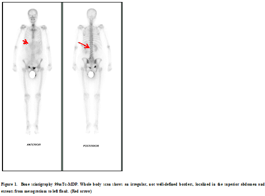
Following imageology malignancy suspicion, bone scintigraphy with 99mTc-MDP is performed without metastasis evidence. However, extraosseous uptake in the abdominal area draws attention, delimiting a substantial mass in this area (Figure 1).
Figure 1: Bone scintigraphy 99mTc-MDP. Whole body scan shows an irregular, not well-defined borders, localized in the superior abdomen and extents from mesogratium to left flank. (Red arrow)
Due to this reason, she is taken to surgical resection of the abdominal mass, finding a retroperitoneal sarcomatous tumor compromising vascular structures of the midline and peritoneal seeding, being able to resect only 60% of the tumor lesion.
Pathology confirmed dedifferentiated liposarcoma (the dedifferentiated component is non-lipogenic) with a high-grade retroperitoneal myxofibrosarcoma pattern associated with significant necrosis areas. Systemic therapy with Gemcitabine + Docetaxel was started while waiting to complete the five cycles and assess evolution.
Discussion
We present a case of a woman in her sixth decade of life with the presence of a giant retroperitoneal mass, which is identified as an extraskeletal lesion with uptake in bone scintigraphy with 99mTc-MDP. The patient underwent partial surgical resection, and histopathology results showed a dedifferentiated liposarcoma with significantly associated necrosis.
Sarcomas are uncommon malignant mesenchymal neoplasia (<1% from all malignant tumors) that have their origin in bone and soft tissues, including a variety of histopathological variants [2]. Within soft tissue sarcomas, the liposarcoma corresponds to the most frequent histopathological type; it originates from cells in the mesoderm and adipose tissue, corresponding to 9-18% from all cases and 10-20% from all primary retroperitoneal malignant tumors [3]. From all retroperitoneal sarcomas, 35% arise from perirenal adipose tissue [3]. It affects men more than women between the fourth and fifth decade of life [4]. The capacity of distant metastasis depends on the grade of histopathological differentiation, thus being the most relevant characteristic [5]. These tumors are prone to dedifferentiate regarding their location, which results in a late diagnosis and time for the tumor to progress and advance [5].
Most of the cases are asymptomatic until the tumor produces a compressive effect, manifesting as a palpable, usually painless (78% of the cases) abdominal mass with an average size of 20-25 cm, thus receiving the name of giant liposarcoma [6]. These findings were present in the initial clinical presentation of our patient.
For its initial diagnosis, a contrast computerized tomography (CT) should be performed. A large, encapsulated mass corresponds to fat and soft tissue presence in the tumor with different attenuation values within [6]. A magnetic resonance image (MRI) is the gold standard for this type of tumor and its correct differential diagnosis [6].
Although a bone scintigraphy image is not considered a diagnostic method, we may find uptake from the radiotracer in this tumors. After the initial injection of MDP, the radiopharmaceutical will freely diffuse into the extravascular fluid; any pathological process that augments the proportion of the radiotracer in the extravascular fluid will, in turn, allow visualization of soft tissues that take in the radiotracer, with its corresponding uptake show in the image [11].An altered local biodistribution of the radiotracer and the extracellular fluid, an increase of the regional blood flow, capillary permeability alteration, neovascularization of the tumor, and the alteration of the sympathetic tone that in turn opens new vascular beds, comprehend a series of events that explain the uptake of the radiopharmaceutical by the tumor [1].
Multiple studies suggest that tumor factors such as calcification or necrosis are additional findings presented in this type of extraskeletal uptake from the radiotracer, probably because of secondary changes present in the mitochondria endoplasmic reticulum due to necrosis [7]. After this happens, both of the mentioned organelles become vacuoles that accumulate calcium salts and minerals that result from cell degradation, leading to a dystrophic calcification, thus allowing a suitable environment for the radiopharmaceutical uptake and its corresponding image in a diphosphonate bone scintigraphy [7].
Various medical specialties must accomplish the treatment of this type of tumor. For localized tumors, surgery with negative margins is the preferred treatment, presenting local recurrence in 40-80% of low-grade tumors cases.6 Neoadjuvant radiation therapy is reserved for high-grade or unresectable tumors, while post-operatory chemotherapy is indicated in patients with advanced or undifferentiated tumors, although no benefit has been proven. Complete resection with negative margins is the most important prognostic factor [6].
Conclusion
Liposarcoma is an infrequent malignant mesenchymal neoplasia that usually occurs between the fourth and fifth decade of life, accounting for approximately 10% of all retroperitoneal tumors, with a typical clinical presentation of a rather big painless abdominal mass, which may be observed in non-indicated imaging modalities, such as 99mTc-MDP bone scintigraphy, with an unusual uptake of a radiotracer that has its main biodistribution is in bone, given the vascular changes, necrosis and dystrophic calcification that occurs in a soft tissue tumor.
References
- Peller PJ, Ho VB, Kransdorf MJ. Extraosseous Tc-99m MDP uptake: a pathophysiologic approach. RadioGraphics. 13, 715-734 (1993).
- Calleja MC, Hernández FJ, López C et al. Subtipos histológicos de liposarcoma: presentación de cuatro casos. Anales de Medicina Interna. 24, (2007).
- Reyna E, Suarez I, Prieto J et al. Liposarcoma retroperitoneal gigante. Reporte de caso. Avances en Biomedicina. 4, 38-42 (2015).
- Foneron A, Vitagliano G, Sánchez-Salas R et al. Mixoliposarcoma perirenal: comunicación de un caso resuelto por vía laparoscópica. Archivos Españoles de Urología (Ed. impresa). 60, (2007).
- Vadillo A, Cornelio G, Méndez M. Liposarcoma retroperitoneal gigante. Acta Médica Grupo Ángeles. 17, 81-83 (2019).
- Liu S, Xie J, Yu F et al. 99mTc-Methylene Diphosphonate Uptake in Soft Tissue Tumors on Bone Scintigraphy Differs Between Pediatric and Adult Patients and Is Correlated with Tumor Differentiation. Cancer Management and Research. 12, 2449-2457 (2020).
Indexed at, Google Scholar, Crossref
Indexed at, Google Scholar, Crossref


