Research Article - Imaging in Medicine (2017) Volume 9, Issue 6
Granular cell tumour of the breast - diagnostic radiology case study
Chandra Segran Shrina Devi, Arasaratnam Shantini & Mohammed Hussein Muna*Faculty of Medicine, Mahsa University, Malaysia
- Corresponding Author:
- Mohammed
Hussein Muna
Faculty of Medicine, Mahsa University, Malaysia
E-mail: dr.mona-mm@mahsa.edu.my
Abstract
A 53 year old female patient presented with a suspicious palpable breast mass with a clinical and radiological suspicion of malignancy. Histological evaluation, however, revealed a rare benign neoplasm. Granular cell tumor is a rare neoplasm that may occur in any anatomical part of the body, most commonly the tongue. GCT of the breast is commonly a benign neoplasm. However, clinically and radiologically it can mimic breast malignancy. It commonly presents as a painless palpable lump while the sonogram and mammogram are variable. FNA or core needle biopsy is diagnostic and wide local excision is the choice of management.
Keywords
granular cell tumor ▪ breast cancer ▪ benign neoplasm ▪ sonogram ▪ mammogram ▪ pathology
Introduction
Granular cell tumor (GCT) is a rare neoplasm that may occur in any anatomical part of the body, most commonly within the oral cavity, the tongue. The first reported case of granular cell tumor involving the tongue was in 1926 by Abrikosoff who proposed the possible origin of GCT from striated muscle cells and he termed this lesion as myoblastoma. However, the histogenesis of this tumor is controversial and currently, the most accepted theory is that it arises from Schwann cell. GCT is commonly benign, however malignant GCT have been reported and comprises less than 2% of GCTs. There have been cases reported of breast GCT synchronous with infiltrating ductal carcinoma. GCT is commonly benign, however malignant GCT have been reported and comprises less than 2% of GCTs . There have been cases reported of breast GCT synchronous with infiltrating ductal carcinoma.
Commonly GCT of the breast is seen in women and rarely in the male breast. It can present as a solitary lump or multi-centric within the breast or other parts of the body.
GCT is commonly benign, however malignant GCT have been reported and comprises less than 2% of GCTs. There have been cases reported of breast GCT synchronous with infiltrating ductal carcinoma .
GCT of the breast accounts for 5-6% of all GCT, approximately 1 per 1,000 cases of breast cancer. Most cases are found in the middle-aged, premenopausal women and more common in African Americans ethnicity.
Clinically, GCT presents as a painless palpable breast mass with a firm to a hard consistency that is common in the upper inner quadrant.
FNAC and core needle biopsy are always diagnostic.
The radiological features are variable, many radiological features are suggestive of breast cancer, that can redirect the management towards unnecessary radical surgical mastectomy, where wide margin excisional surgical removal is the effective management for begnin GCT where no axillary lymph nodes involvement.
Increasing the level of knowledge and awareness to Include GCT in the differential diagnosis of and suspicious painless breast lump will redirect the management pathways and will have great impacts on health care system, socioeconomic aspects , and patient safety.
Case history
A 53 year old female patient presented with a suspicious palpable breast mass with a clinical and radiological suspicion of malignancy. Histological evaluation, however, revealed a rare benign neoplasm.
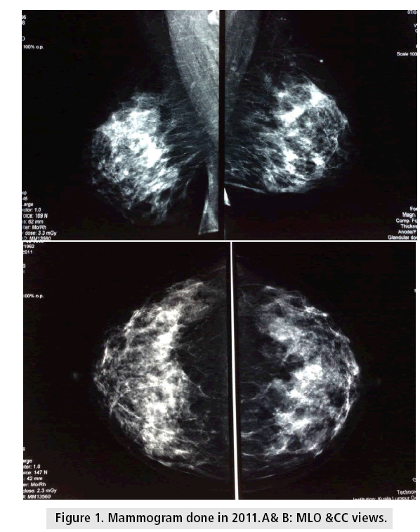
Referral to Hospital Kuala Lumpur (HKL) from a private center following a screening mammogram which revealed a left breast lump. Upon further history taking and clinical examination revealed that patient was previously under surgical clinic follow up for a then nonpalpable left breast lump since 2001 [1]. The lesion was detected on the mammogram and biopsied sometime in 2004 by the surgical team. The patient was told that the lesion was benign in nature. The patient continued to follow up and then defaulted sometime in 2011.
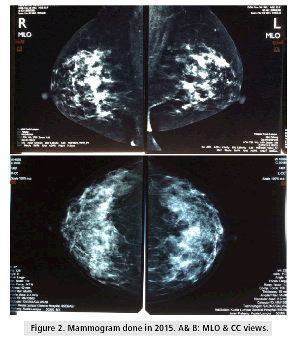
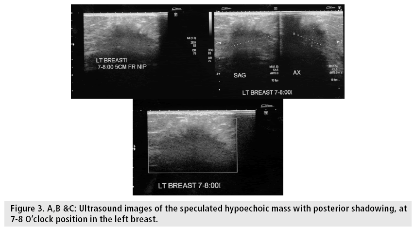
Following the referral back to HKL, the patient underwent mammogram and ultrasound examination. The mammogram and ultrasound results in 2011 were no mass on both mammogram and ultrasound (BIRADS 1) while in 2015 no mass detected on the mammogram, however by ultrasound, an irregular hypoechoic lesion noted in the left breast at 7-8 O’ clock position (BI-RADS 4) [2].
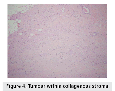
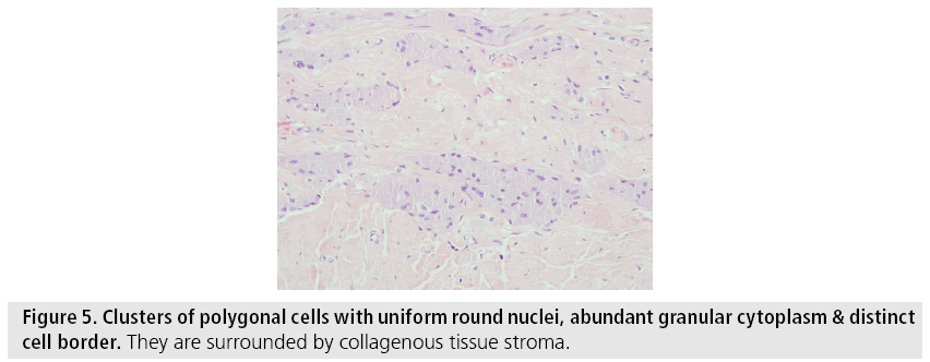
Intraoperative findings revealed a stony hard lump measuring approximately 2 × 2 cm with no underlying pectoralis muscle involvement [3]. The histopathology (HPE) report was granular cell tumor, inadequate excision [4] (FIGURES 1-5 and TABLES 1 and 2).
Figure 5: Clusters of polygonal cells with uniform round nuclei, abundant granular cytoplasm & distinct cell border. They are surrounded by collagenous tissue stroma.
| Clinical Correlation | |||
|---|---|---|---|
| Clinical presentation (2011) | Right | Left | Remarks |
| a. Lump in breast | - | - | |
| b. Nipple discharge | - | - | |
| c. Nipple reaction | - | - | |
| d. Discomfort/tenderness/pain | - | - | |
| e. Axillary node swelling | - | - | |
| Relevant Personal History | Yes | No | Remarks |
|
/ | ||
|
/ | ||
|
/ | ||
|
/ | ||
|
/ | 2002-2005 | |
|
|||
|
A
A
A
|
||
| Radiological Findings | |||
| Remarks | |||
| Mammogram findings | Bilateral dense breast with scattered microcalcifications | ||
| Mass | No dominant mass bilaterally | ||
| USG | Not done | ||
| Impression | No mammographic evidence of malignancy | ||
| Follow up | Routine | ||
Table 1: Mammogram report in 2011.
| Clinical Correlation | |||
|---|---|---|---|
| Clinical presentation (2015) | Right | Left | Remarks |
| a. Lump in breast | - | / | |
| b. Nipple discharge | - | - | |
| c. Nipple reaction | - | - | |
| d. Discomfort/tenderness/pain | - | - | |
| e. Axillary node swelling | - | - | |
| Relevant Personal History | Yes | No | Remarks |
| Personal history of breast cancer | / | ||
| First degree relatives with breast cancer | / | ||
| Previous breast surgery or trauma | / | ||
| HRT | / | ||
| Birth control pills | / | 2002-2005 | |
| Breast fed children | / | ||
| Any previous MMG | / | ||
| Radiological Findings | |||
| Remarks | |||
| Mammogram findings | Bilateral dense breast with scattered microcalcifications | ||
| Mass | No dominant mass bilaterally | ||
| USG | Right breast – NAD Left Breast – an irregular hypoechoic lesion at 7-8 O’clock (Lower inner quadrant) measuring 1.5 × 2.1 × 3.4 cm) |
||
| Impression | Suspicious left 7-8 O’clock breast lesion, BIRADS 4 | ||
| Follow up | US guided biopsy March 2015 (5 attempts made, however only 1 sample attained. Decision made for excision biopsy | ||
Table 2: Mammogram report in 2015.
Discussion
Granular cell tumor (GCT) is a rare neoplasm that may occur in any anatomical part of the body, most commonly within the oral cavity, the tongue. The first reported case of granular cell tumor involving the tongue was in 1926 by Abrikosoff who proposed the possible origin of GCT from striated muscle cells and he termed this lesion as myoblastoma [5].
However, the histogenesis of this tumor is controversial and currently, the most accepted theory is that it arises from Schwann cell.
GCT is commonly benign, however malignant GCT have been reported and comprises less than 2% of GCTs [6]. There have been cases reported of breast GCT synchronous with infiltrating ductal carcinoma [7].
GCT of the breast accounts for 5-6% of all GCT, approximately 1 per 1,000 cases of breast cancer [8]. Most cases are found in the middle-aged, premenopausal women and more common in African Americans ethnicity [9].
Commonly GCT of the breast is seen in women and rarely in the male breast. It can present as a solitary lump or multi-centric within the breast or other parts of the body [10].
Presentation
Clinically, GCT presents as a painless palpable breast mass with a firm to a hard consistency that is common in the upper inner quadrant [11]. GCT can also cause skin retraction and ulceration, due to its infiltrating growth pattern and close apposition to the pectoralis muscle and fascia.
The radiologic features of GCT of the breast are variable.
By ultrasound, it could be seen as a solid, hypoechoic, poorly marginated mass, associated with posterior acoustic shadowing or as a more benign- appearing well-circumscribed mass with posterior acoustic enhancement. In Mammogram it may present as a round or smooth lobulated, well-circumscribed configuration or as an ill-defined spiculated mass. Microcalcifications are not present [2].
In a case series by Irshad et al, the sonographic and mammographic appearances of GCT were also found to be variable [12]. The common sonographic features of GCT were a hypoechoic lesion and taller than wider (height to width ratio>1). While the mammographic findings of the tumor showed speculations with no calcification [13].
These findings on both sonography and mammography show that GCT mimics breast malignancy.
Investigation
FNAC and core needle biopsy are always diagnostic. However, in our patient biopsy attempt failed as the mass was too hard to attain good sample. Hence a wide local excision of the tumor was done.
Macroscopically, GCT appears firm to hard mass and grayish to yellow in color in its cross section [14]. Microscopically, it shows the abundance of eosinophilic cytoplasm that has a granular appearance caused by the accumulation of mitochondria, secretory granules and lysosomes [15].
There have been case reports where MRI and PET were carried out for a pre-operative diagnosis of GCT. Dynamic MRI showed strong enhancing lesion with peripheral rim enhancement and no washout phenomenon.
While PET, visual analysis did not show a focal enhanced tracer accumulation and quantitative image analysis (FDG uptake by tumor) using average SUV revealed less than the threshold SUV of 2.5, which was used to differentiate benign and malignant lesion [14].
However neither of these imaging was done in our patient, because the patient underwent surgery.
Based on a study conducted by Papalas et al. [16] it was noted that the risk of disease progression was low. Despite resection of the tumor with positive margins, there was no recurrence of tumor up to 7years post resection. However, long-term follow-up of patients is recommended as recurrence after 10years post resections have been reported [16].
Management
Wide local excision with clear margin is the surgery of choice for benign GCT. Axillary clearance is not necessary as nodal involvement is rare. Chemotherapy and radiotherapy have no role in benign GCT of the breast. However, the same can’t be said for its malignant counterpart. Malignant GCT has been reported to be progressive with metastasis and thus may require chemo or radiotherapy in its management [17].
Conclusion
In conclusion, GCT of the breast is a rare benign neoplasm, which appears to present clinically and have radiological features that are more aggressive than it truly is. Awareness of its many variable presentations will benefit both the radiologists and surgeons.
References
- Damiani S, Koerner FC, Dickersin RG et al. Granular cell tumor of the breast. Virchow. Archiv. A. Pathol. Anat. 420, 219-226 (1992).
- Irshad A, Pope TL, Ackerman SJ et al. Characterization of sonographic and mammographic features of granular cell tumors of the breast and estimation of their incidence. J. Ultrasound. Med. 27, 467-475 (2008).
- Patel A, Lefemine V, Yousuf SM et al. Granular cell tumor of the pectoral muscle mimicking breast cancer. Cases. J. 1, 142 (2008).
- Lack EE, Worsham GF, Callihan et al. Granular cell tumor: A clinicopathologic study of 110 patients. J. Surg. Oncol. 13, 301-316 (1980).
- Abrikossoff A, Über M, Ausgehend VQ et al. About myomas. Virchows. Arch. Pathol. Anat. 260, 215-233 (1926).
- Adeniran A, Al-Ahmadie H, Mahoney MC et al. Granular cell tumor of the breast: A series of 17 cases and review of the literature. Breast. J. 10, 528-530 (2004).
- Bonito MD, et al. Coexistence of granular cell tumor and invasive ductal breast cancer in contralateral breasts: A case report. Int. J. Mol. Sci. 15, 13166-13171 (2014).
- Feder JM. Unusual breast lesions: Radiologic-pathologic correlation. Radiographics. 19, 11-26 (1999).
- Chetty R, Kalan MR. Malignant granular cell tumor of the breast. J. Surg. Oncol. 49, 135 (1992).
- Gogas J, Markopoulos C, Kouskos E et al. Granular cell tumor of the breast: a rare lesion resembling breast cancer. Eur. J. Gynaecol. Oncol. 23, 333-334 (2002).
- Khansur T, Balducci L, Tavassoli M. Granular cell tumor: clinical spectrum of the benign and malignant entity. Cancer. 60, 220-222 (1987).
- Yang WT, Edeiken MB, Sneige N et al. Sonographic and mammographic appearances of granular cell tumors of the breast with pathological correlation. J. Clin. Ultrasound. 34, 153 (2006).
- Gordon AB, Fisher C, Palmer B et al. Granular cell tumor of the breast. Eur. J. Surg.Oncol. 1, 269-273 (1985).
- Hoess C. FDG PET evaluation of granular cell tumor of the breast. J. Nucl. Med. 39, 1398-1401 (1998).
- Filipovski V, Banev S, Janevska V. Granular cell tumor of the breast: A case report and review of the literature. Cases. J. 2, 51-85 (2009).
- Papalas JA. Recurrence risk and margin status in granular cell tumors of the breast: A clinicopathologic study of 13 patients. Arch. Pathol. Lab. Med. 135, 890-895 (2011).
- Tawfiq K. Granular cell tumor - Clinical spectrum of the benign and malignant entity. Cancer. 60, 220-222 (1978).
