Clinical images - Imaging in Medicine (2018) Volume 10, Issue 6
Imaging characteristics of mature cystic teratoma
Sindhu Kumar*Department of Radiology, University of Virginia Medical Center, Virginia 22901, USA
- Corresponding Author:
- Sindhu Kumar
Department of Radiology
University of Virginia Medical Center
Virginia 22901, USA
E-mail: sindhucumar@gmail.com
Abstract
Keywords
computed tomography, mature cystic teratoma
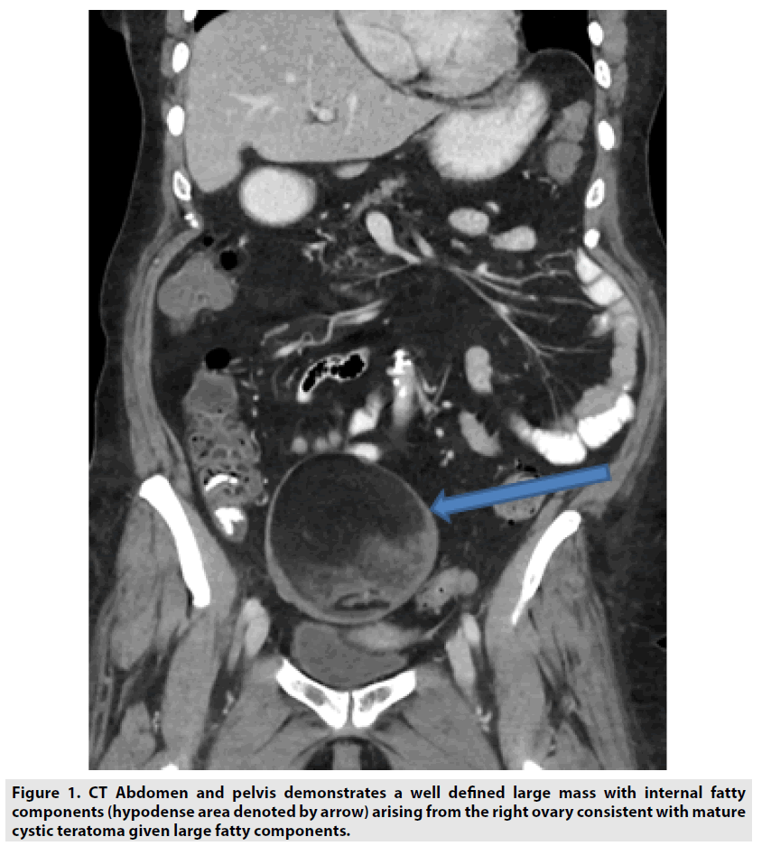
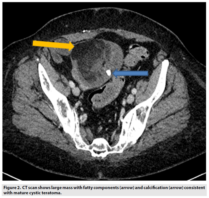
Mature cystic teratoma is one of the most common, slow growing ovarian neoplasm found in a wide range of patient population. They are usually composed of elements two out of three germ cell layers like ectoderm, endoderm or mesoderm. It can be detected by ultrasound or CT scan with characteristic imaging findings. Presence of abundant fat, calcification or dentals components are often seen. Large lesions tend to have malignant potential (FIGURES 1 and 2).


