Research Article - Interventional Cardiology (2022)
Incidence of congenital anomalies of the coronary arteries in embalmed cadavers
- Corresponding Author:
- Santiago Cubas
Departamento de Anatomía,
Facultad de Medicina,
Universidad de la República,
Montevideo,
Uruguay,
E-mail: cubassantiago@hotmail.com
Received date: 30-Mar-2022, Manuscript No. FMIC-22-58956; Editor assigned: 01-Apr-2022, PreQC No. FMIC-22-58956 (PQ); Reviewed date: 18- Apr-2022, QC No. FMIC-22-58956; Revised date: 25-Apr-2022, Manuscript No. FMIC-22-58956 (R); Published date: 02-May-2022, DOI: 10.37532/1755- 5310.2022.14(S9).224
Abstract
Introduction: Coronary anomalies, whose incidence is 0.17% to 1.5%, are relevant since they can debut as sudden cardiac death and can determine technical difficulties in interventional procedures such as coronary angiography. This prompted the following study, whose objective is to determine the incidence of anomalies and anatomical variants in the origin and proximal course of the coronary arteries, in a cadaveric population.
Materials and methods: 81 hearts were dissected from adult cadavers, of both sexes, previously fixed and preserved in a 10% formaldehyde-based solution. Once the heart was released, the identification and subsequent dissection of the coronary arteries and coronary ostia were carried out. The following data were recorded: Number of ostiums, aortic sinus where said ostiums are located, artery that gives origin, route and direction of the same. The data were recorded in tables for later analysis.
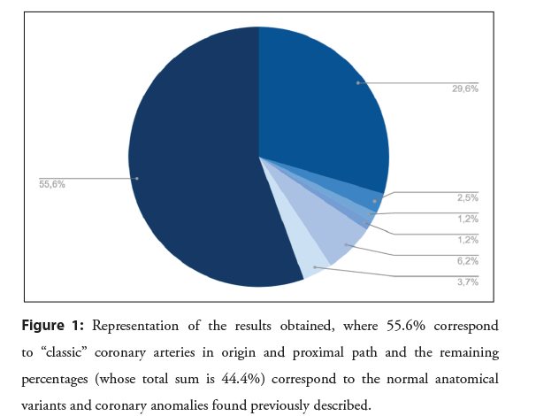
Results: Of the total of 81 dissected hearts, 45 (55.6%) presented “classic” coronary arteries in origin and proximal path and 36 (44.4%) presented normal anatomical variants and coronary anomalies.
Conclusion: Knowledge of coronary anomalies is of the utmost importance, given that between 20% to 90% present with sudden cardiac death, given that when it comes to performing interventional procedures, their ignorance may determine an increase in the duration of the procedures with greater contrast input and radiation exposure for the patient, and in cardiac surgery during de infusion of cardioplegia directly by coronary ostiums.
Keywords
Coronary anomalies • Sudden death • Coronary angiography
Introduction
Coronary anomalies are defined as variations in their origin, trajectory, intrinsic anatomy, anastomosis and/or termination of the coronary arteries [1]. Initially, they were considered a benign finding of Coronary Angiography (CACG) [2-5] or were diagnosed incidentally during autopsy [6]. Their incidence according to the literature is 0.17 to 1.5% [1-4,6-8], its importance is that they can present as sudden cardiac death [1,3] and they can determine technical difficulties in interventional procedures such as CACG, exposing the patient to higher doses of intracoronary contrast and longer exposure time to radiation [1,8,9], and in cardiac surgery difficulties in myocardium protection with the infusion of cardioplegia, in surgery of aorta, aortic valve and Coronary Artery Bypass Graft (CABG).
The increased number of diagnoses in recent years were mainly due to greater medical knowledge, the continuous development of imaging techniques and their greater availability [6,10]
The objective of this study is to determine the incidence of anomalies and anatomical variants in their origin and proximal trajectory of the coronary arteries in the cadaveric population of the Departamento de Anatomía, Facultad de Medicina, Universidad de la República, Montevideo, Uruguay.
Materials and Methods
The present work is a descriptive anatomical study, of a crosssectional observational type, for which 81 hearts from adult cadavers of both sexes were dissected, previously fixed and preserved in 10% formaldehyde-based solution, belonging to the Departamento de Anatomía, Facultad de Medicina, Universidad de la República. The average age of the donors was 55 to 65 years.
The dissection started with a bilateral thoracotomy following the anterior axillary line and later, sectioning the superior and inferior vena cava and the supracardiac vessels, the cardiopulmonary block was excised. Both pulmonary hilum and the rest of the large supracardiac vessels were dissected. Concomitantly, an inverted “T” pericardiotomy was performed and the intrapericardial pulmonary veins were sectioned, thus freeing the heart.
Once the heart was released, the coronary arteries and coronary ostia were identified and later dissected.
The following data were recorded: Number of ostiums, aortic sinus where said ostiums are located, artery that gives origin, route and direction of the same. The data were recorded in tables for later analysis.
A bibliographic search was carried out in the following electronic databases: Medline, Portal Timbó, Cochrane and Pubmed; using as keywords: “coronary arteries”, “anomalous origin”, “congenital anomaly” in combination with the boolean operators “AND”, “OR”. The collected articles were used for the discussion and analysis of our findings and are cited in the corresponding section.
Results
Of the total of 81 dissected hearts, the following results were obtained: 45 (55.6%) presented “classic” coronary arteries in origin and proximal course and 36 (44.4%) presented normal anatomical variants and coronary anomalies.
Regarding the total of normal anatomical variants and coronary anomalies, the results were as follows: 24 (29.6%) presented trifurcation of the left coronary artery; 5 (6.2%) had an independent origin of the artery of the conus arteriosus (conal artery) in the right aortic sinus; 3 (3.7%) presented trifurcation of the left coronary artery and an independent origin of the conal artery in the right aortic sinus; 2 (2.5%) presented quadrifurcation of the left coronary artery; 1 (1.2%) presented independent origins of the anterior interventricular and circumflex arteries; 1 (1.2%) presented origin of the right coronary artery in the non-coronary aortic sinus (Figure 1).
Discussion
Taking into consideration that the coronary arteries are defined by the cardiac territory of vascularization, the right coronary artery is the one that irrigates the three right and lower quarters of the right ventricle, the right half of the inferior face of the left ventricle and the posterior third of the interventricular septum. The left coronary is responsible for supplying the left third of the anterior wall of the right ventricle, the left ventricle (which is not supplied by the right coronary artery), and the anterior two-thirds of the interventricular septum [11,12].
We consider the following distribution as classic coronary anatomy; the right coronary artery originates in the right aortic sinus through a single ostium. It courses down the right coronary groove, then it takes the posterior face of the heart to end, in most cases, as the posterior interventricular artery, which will run through the homonymous groove. The left coronary artery originates in the left aortic sinus through a single ostium. It courses to the left, between the pulmonary trunk and the left atrial appendage, then to down and forward. After a short journey, this artery bifurcates into the anterior interventricular artery, which runs through the homonymous groove, and the circumflex artery, which runs through the left portion of the coronary groove.
The difference between a coronary anomaly and a normal anatomical variant depends on its incidence in the general population, less than 1.5% is considered a coronary anomaly and greater than 1.5% is a normal anatomical variant. From the results found, the trifurcation of the left coronary artery, the independent origin of the conal artery in the right aortic sinus, the trifurcation of the left coronary artery together with an independent origin of the conal artery and the quadrifurcation of the left coronary artery are normal anatomical variants. Whereas the independent origin of the anterior interventricular and circumflex arteries and the origin of the right coronary artery in the non-coronary aortic sinus correspond to coronary anomalies [1,2,6,10].
Regarding normal anatomical variants, the percentage found in our study could be elevated due to the low number of hearts studied, one of the limitations of our work. These variants have no clinical relevance and can even be considered a protective factor against coronary events, such as an independent origin of the conal artery, since it would not be affected by the compromise of the right coronary artery and therefore, less myocardium would suffer ischemia.
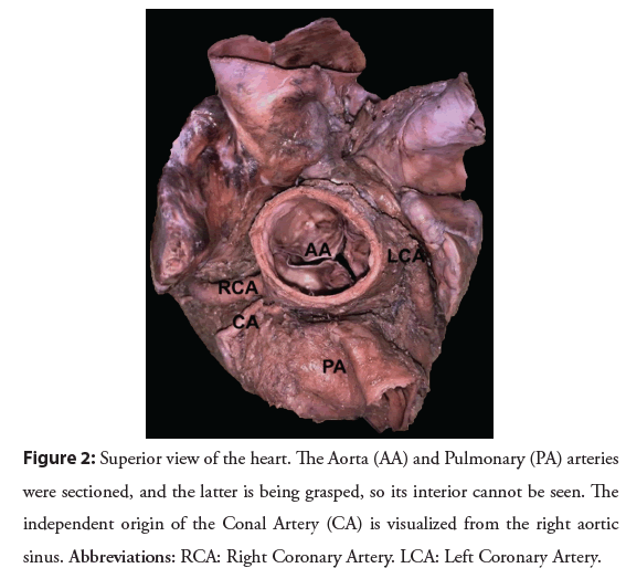
The conal artery, frequently poorly described, presents five patterns of origin according to [13], Type A where it originates as a branch of the right coronary artery, Type B where it originates from a common coronary ostium with the right coronary artery, Type C, pattern found on our dissections (Figure 2). Where it originates independently of the right aortic sinus, Type D where multiple conal arteries originate as separate branches from the right coronary artery and Type E where the conal artery originates as a branch from a right ventricular artery or acute marginal artery. This artery can vascularize a large myocardial territory through anastomotic circuits such as the Vieussens arterial ring.
Figure 2: Superior view of the heart. The Aorta (AA) and Pulmonary (PA) arteries were sectioned, and the latter is being grasped, so its interior cannot be seen. The independent origin of the Conal Artery (CA) is visualized from the right aortic sinus. Abbreviations: RCA: Right Coronary Artery. LCA: Left Coronary Artery.
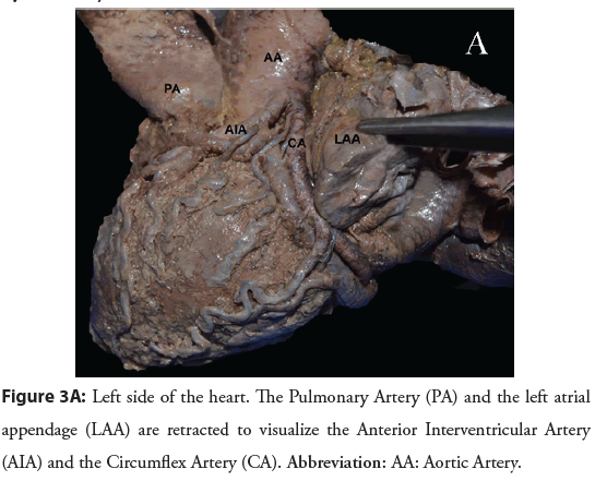
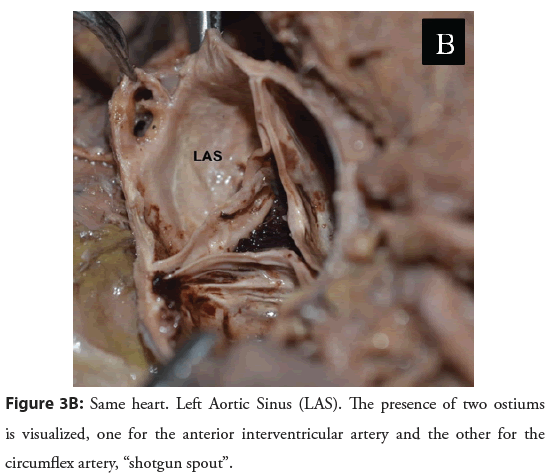
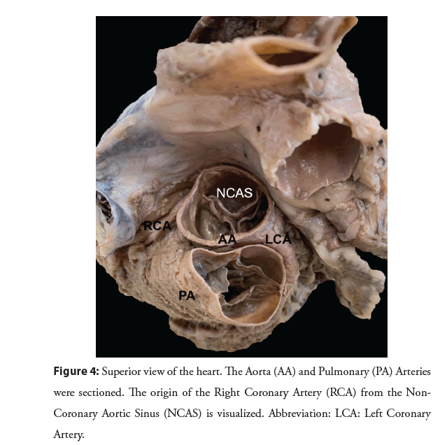
Regarding coronary anomalies, these were found in two of the total of hearts studied, so their incidence was 2.4%, where 1.2% corresponds to the independent origin of the anterior interventricular and circumflex arteries (Figures 3A and 3B) and 1.2% corresponds to the origin of the right coronary artery in the non-coronary aortic sinus (Figure 4). These percentages are higher than those found in the literature, where according to [2] the incidence, by imaging methods, of the independent origin of the anterior interventricular and circumflex arteries is 0.02% and according to [6] the incidence, by imaging methods, of the origin of a coronary artery in the non-coronary aortic sinus is 0.35%. This last percentage is for both coronary arteries (right and left), but it’s an understanding that the origin of the left coronary artery in the non-coronary aortic sinus is extremely rare. The independent origin of the anterior interventricular and circumflex arteries can be considered as a protective factor, since left coronary artery disease is prevented [1]. On the other wise this type of anomalies can produce problems with myocardium protection when the infusion of cardioplegia during cardiac surgery is infused directly by coronary ostiums.
Figure 4: Superior view of the heart. The Aorta (AA) and Pulmonary (PA) Arteries were sectioned. The origin of the Right Coronary Artery (RCA) from the Non- Coronary Aortic Sinus (NCAS) is visualized. Abbreviation: LCA: Left Coronary Artery.
The clinical presentation of coronary anomalies is variable, from sudden death, acute myocardial infarction, arrhythmias, heart failure, syncope, palpitations, or asymptomatic patients [2- 6,10,14-18,19].
An anomaly to be highlighted due to its clinical importance is the anomalous origin of the left coronary artery from the pulmonary artery or from the right or left pulmonary artery, known as Bland- White-Gerland syndrome, a very common coronary anomaly in children, whose approximate global incidence is 1 in 300,000 children [3,7].
Another anomaly of great clinical relevance is the interarterial course of the coronary arteries, since there is a great risk of compression between the aorta and the pulmonary artery; this path is frequently seen when the coronary artery originates from the opposite aortic sinus [2, 3,7,10,14].
Finally, certain coronary anomalies are considered benign, such as the origin of the right coronary artery from the non-coronary aortic sinus [1].
Diagnostic confirmation of coronary anomalies is carried out by imaging methods, being the CACG, computed tomography angiography (CT-angiography) and magnetic resonance angiography (MRI-angiography) the methods of choice. The disadvantage of CACG is that it provides two-dimensional images and that it is an invasive method, which involves arterial cannulation, uses ionizing radiation and contrast. In contrast to this, CT-angiography and MRI-angiography provide threedimensional images, are not invasive methods and present fewer complications [14,17].
Conclusion
Knowledge of coronary anomalies is of the utmost importance, mainly because they can clinically present as sudden cardiac death, a presentation that occurs in 20 to 90% of coronary anomalies and an etiological cause in 11% to 12% of sudden cardiac deaths. We also emphasize its importance when performing interventional procedures, since its ignorance may determine an increase in the duration of the procedures with greater contrast input and radiation exposure for the patient, and in cardiac surgery during de infusion of cardioplegia directly by coronary ostiums.
Acknowledgements
All the authors are deeply grateful to those who donated their bodies for teaching and research to the Departamento de Anatomía, Facultad de Medicina, Universidad de la República.
References
- Kayalar N, Burkhart HM, Dearani JA, et al. Congenital coronary anomalies and surgical treatment. Congenit Heart Dis. 4(4): 239-251 (2009).
[CrossRef] [Google Scholar] [PubMed]
- Barriales R, Morís C, López A, et al. Adult congenital anomalies of the coronary arteries described over 31 years of angiographic studies in the Asturias Principality: main angiographic and clinical characteristics. Rev Esp Cardiol. 54(3): 269-281 (2001).
[CrossRef] [Google Scholar] [PubMed]
- Luchessi E, Calenta C, Krischmann D, et al. Anomalías coronarias congénitas y aterosclerosis: Nuevo factor de riesgo? Rev Fed Arg Cardiol. 40: 407-409 (2011).
- Palmieri V, Gervasi S, Bianco M, et al. Anomalous origin of coronary arteries from the “wrong” sinus in athletes: Diagnosis and management strategies. Int J Cardiol. 252: 13-20 (2017).
[CrossRef] [Google Scholar] [PubMed]
- Sinha SK, Aggarwal P, Razi M, et al. Unduly long left main (79 mm) coronary artery arising from right coronary sinus in a 64-year-old diabetic man. BMJ Case Rep. 12(7): e229815 (2019).
[CrossRef] [Google Scholar] [PubMed]
- Hosoda R, Masuoka A, Nagase H, et al. Anomalous aortic origin of a coronary artery in two children. Asian Cardiovasc Thorac Ann. 28(1): 55-58 (2020).
[CrossRef] [Google Scholar] [PubMed]
- Hlavacek A, Loukas M, Spicer D, et al. Anomalous origin and course of the coronary arteries. Cardiol Young. 20(S3): 20-25 (2010).
[CrossRef] [Google Scholar] [PubMed]
- Tyczynski P, Kukula K, Pietrasik A, et al. Anomalous origin of culprit coronary arteries in acute coronary syndromes. Cardiol J. 25(6): 683-690 (2018).
[CrossRef] [Google Scholar] [PubMed]
- Loukas M, Andall RG, Khan AZ, et al. The clinical anatomy of high take-off coronary arteries. Clin Anat. 29(3): 408-419 (2016).
[CrossRef] [Google Scholar] [PubMed]
- Vinnakota A, Stewart RD, Najm H, et al. Anomalous aortic origin of the coronary arteries: A Novel Unroofing technique in an adult cohort. Ann Thorac Surg, 107: 823-828 (2019).
[CrossRef] [Google Scholar] [PubMed]
- Latarjet M, Ruiz Liard A. Anatomía humana. 5th ed. Buenos Aires; Madrid [etc.]: Editorial Médica Panamericana, pp. 860-885 (2019).
- Kouchoukos NT, Blackstone EH, Hanley FL, et al. Cardiac Surgery. 4th ed. United States: Elsevier Saunders, pp. 1-66 (2013).
- Loukas M, Patel S, Cesmebasi A, et al. The clinical anatomy of the conal artery. Clin Anat. 29(3): 371-379 (2016).
[CrossRef] [Google Scholar] [PubMed]
- Lorenzoni G, Merella P, Viola G, et al. Anomalous origin of right coronary artery from left sinus of valsalva. J Invasive Cardiol. 31(9): E279 279.
[Google Scholar] [PubMed]
- Pawale A, Takahashi M, Seetharam K, et al. Minimally invasive direct coronary artery bypass for the management of anomalous left coronary artery from the right coronary sinus. Heart Surg Forum. 21(4): 239-241 (2018).
[CrossRef] [Google Scholar] [PubMed]
- Reddy RC, Takahashi M, Beckles DL, et al. Anomalous right coronary artery from the left sinus: A minimally invasive approach. Eur J Cardiothorac Surg, 41(2): 287-290 (2012).
[CrossRef] [Google Scholar] [PubMed]
- Bachini JP, Amodio A, Guzmán R, et al. Nacimiento anómalo de la arteria coronaria izquierda desde la arteria pulmonar, síndrome de ALCAPA. Primer reporte de caso en Uruguay. Rev Urug Cardiol. 34(2): 213-217 (2019).
- Finocchiaro G, Behr ER, Tanzarella G, et al. Anomalous coronary artery origin and sudden cardiac death: Clinical and pathological insights from a national pathology registry. JACC Clin Electrophysiol. 5(4): 516-522 (2019).
[CrossRef] [Google Scholar] (All versions) [PubMed]
- Kamperidis V, Karamitsos TD, Pappa Z, et al. ALCAPA syndrome and risk of sudden death in young people. QJM. 112(4): 291-292 (2019).
[CrossRef] [Google Scholar] [PubMed]
