Research Article - Pharmaceutical Bioprocessing (2018) Volume 6, Issue 3
Isoginkgetin inhibits lipopolysaccharide induced PI3K/AKT pathway through miRNA210 in vascular endothelial cells
- *Corresponding Author:
- Lihua Han
Henan University of Traditional Chinese Medicine
Zhengzhou, 450008, China
E-mail: xeaxeng98@163.com
Abstract
Objective: To investigate whether isoginkgetin can inhibit lipopolysaccharide induced PI3K/ AKT pathway through miRNA210 in vascular endothelial cells.
Methods: The experiment was used isoginkgetin 20 uM to treat lipopolysaccharide induced endothelial cells and to observe the protein and gene expression of miRNA210 and phosphatidylinositol 3-kinase (PI3K)/AKT signaling pathways. In addition, the effect of isoginkgetin on the PI3K/AKT signaling pathway was observed after the miRNA210 gene was overexpressed by the virus. The experiment mainly tested by PCR and Western blot to detect the gene and protein expression.
Results: The results showed that isoginkgetin could significantly inhibit the expression of miRNA210, PI3K and Akt in the vascular endothelial cells induced by lipopolysaccharide. The effect of isoginkgetin was significantly inhibited after the use of viral overexpression of miRNA210.
Conclusion: Isoginkgetin mainly inhibits the PI3K/AKT signaling pathway in lipopolysaccharide induced vascular endothelial cells by inhibiting miRNA210. In addition, miRNA210 may play an important role in many PI3K pathway related diseases.
Keywords
isoginkgetin, miR-210, vascular endothelial cells, PI3K, Akt
Introduction
As early as 1960s, scholars began to use ginkgo leaf total flavonoids to study the vasodilator activity and apply it to clinical efficacy. In recent years, the high tide of further research on isoginkgetin has been raised in China. A large number of studies have proved that isoginkgetin has many advantages, such as blood lipid, anti-tumor, anti-inflammatory, anti-radiation, anti-leukemia and so on [1].
MicroRNAs (miRNA) are a class of about 18~25 nucleotides (nt) endogenous, non-coding single-stranded RNA molecules and its sequences are highly conserved among various species with the same regulatory mechanism in different developmental stages [2]. The study has found that [3] miRNA can specifically recognize the 3’untranslated region of the mRNA of target gene to inhibit its translation or promote its degradation with a negative regulatory effect on post-transcriptional level, thereby taking a part in a variety of biological process such as cell proliferation, differentiation, apoptosis, metabolism, hormone secretion, ontogeny and disease development. miRNA is strictly tissue-specific and time-dependent and plays an important regulatory role in development of cardiovascular system as well as disease. Many experiments have shown that [4] miRNA has specific gene expression profiles in cardiovascular disease, indicating that it may become a new biomarker for disease diagnosis, treatment and prognosis evaluation. Therefore, further research on the function of miRNA has become a hot topic in the field of cardiovascular disease.
Among numerous changing miRNAs, the expression of miR-210 is most stable and obvious [5]. Under hypoxic conditions, miR-210 can inhibit the cell proliferation, hinder mitochondrial metabolism for a moderately long time, weaken the capability of DNA damage repair and promote neovascularization, thus playing a unique and important role in hypoxic diseases and pathological process of tumors [6]. Moreover miR-210 plays a prominent effect in ischemic heart and cerebral vascular diseases. It is shown in the related study that [7] in case of animal hippocampus ischemia miR-210 is up regulated and the target gene ephrin-A3 notably down regulated. MiR-210 expression was elevated in animal models of cardiac hypertrophy, heart failure, transient focal cerebral ischemia, lower limb ischemia, and cerebral ischemia injury in which ischemic injury is the main cause of organ failure and to repair the injury, promotion of angiogenesis and restoration of effective blood supply is the key. Studies have shown that [8] phosphatidylinositol 3-kinase (PI3K)-protein kinase B (PKB/Akt) negatively regulates TF expression in endothelial cells and it can be activated by serine 1177 of Akt phosphorylation endothelial nitric oxide synthase (eNOS) to promote the generation of nitric oxide (NO). NO is a potent vasodilator that inhibits cell proliferation as well as platelet aggregation in smooth muscle and TF expression in endothelial cells. However, the regulation pathway and mechanism of miRNA210 on PI3K/AKT signaling pathway in vascular endothelial cells are still unknown. For this reason, we used isoginkgetin to observe whether it can affect the PI3K/AKT signaling pathway by regulating miRNA in lipopolysaccharide induced PI3K/AKT signaling pathway in vascular endothelial cells.
Materials and methods
Materials
Human umbilical vein endothelial cells (HUVE-12) were purchased from Shanghai Meiyan Biotechnology Co., ltd.
Main reagents
Trizol reagent (Shanghai Kemin Biotechnology Co., Ltd.); reverse transcription kit (Hangzhou Connaught Biological Technology Co. Ltd.); PE-labeled II (Shanghai Fanke Biotechnology Co. Ltd.); PI3K antibody (Yi Baikang Biological Polytron Technologies Inc); Akt antibody (Beijing Boosen Biological Technology Co., Ltd.).
Cell culture
1 * 109/ L HUVE-12 cells were inoculated in culture flasks and cultured in the RPMI -1640 culture liquid with 15% FBS followed by being placed in 5% CO2 incubator for culture at 37 with the medium changed the next day. Lipopolysaccharide, Isoisoginkgetin, or overexpression of virus were used to treat cells for 36 h. The cells attached to the wall and sprawled out in the shape of cobblestone; Digestion and passage of 0.25% EDTA trypsin were performed until 80% confluence of cells.
Construction and transfection of recombinant lentiviral vector
The PCR primer suitable for the target sequence was designed. The two synthesized primers were annealed and the target carrier as well as the annealing product were respectively digested by double enzymes. primers of miRNA210 for PCR were sense 5’- GAAAUGAGCUGGUAAAGAATT -3′and antisense: 5’- UUCUUUACCAGCUCAUUUCTT-3.
The digestion products which were purified and recovered were directly ligated followed by transformation of competent cell, then cell clones were firstly identified by PCR with both upstream and downstream primers designed on the carrier, if the PCR identification turned out to be positive, it proved that the target fragment has been connected to purpose vehicle. Then clones with positive result of PCR identification were sequenced and analyzed and the correct fragments in comparison with each other were exactly successfully-constructed plasmid vectors. The endothelial cells were inoculated into 6 pore plates with the density of 3. 0 × 108/ L 24h L before transfection. With the multiplicity of infection (MOI) being 10, 0.75 L virus-containing fluid was added (with the final concentration of 3. 0 × 109 cell /L) and Polybrene was added to each hole with the final concentration of 5 mg / L. at 72 h after transfection, GFP green fluorescence was observed under fluorescence microscope and the transfection efficiency was estimated. The experiment included miR-210 overexpression group and control group.
Detection of changes in expression of miR- 210, PI3K and AKT by real-time PCR
Cells were collected 72 h after transfection and total RNA was extracted with Trizol method. The ratios of A260/A280 and A260/ A280, required to range from 1.8 to 2.0, were measured by nucleic acid determinator and the quality of RNA was assessed by using 1% agarose gel electrophoresis. MiR-210 reverse transcription was performed by stem-loop primers with following reaction conditions: 16°C 30 min, 30°C 30 s, 42°C 30 s, 50°C 1 s, with a total of 60 cycles; at 70°C, 15 min, U6 was taken as the internal reference and common gene expression was detected with GAPDH as the internal reference. The genes were amplified with the method of SYBR Green qPCR and detected by ABI 7500 real-time PCR system with following reaction conditions: 95°C 10 min, 95°C 15 s, 60°C 30 s, with a total of 40 cycles followed by the dissociation curve analysis in the end. Three repeats were set up in each sample with template-free as negative control and relative changes of gene expression were described as “2-ΔΔCt”.
Detection of changes in expression of PI3K and AKT by Western blotting
The endothelial cells were collected. The total protein was extracted and quantified according to the instruction of extraction kit. 30 g protein sample was taken followed by PAGE electrophoresis and transferred to nitrocellulose membrane for 2 h and closed with 5% skimmed milk powder at room temperature for 1 h. I-resist (1: 1000 rat anti human PI3K or AKT) was added and incubated at 37°C for 2 h with TBST buffer washing of membrane, horseradish peroxidase-labeled goat anti rat IgG (1: 3000) was added and incubated at 37°C for 1 h also with TBST buffer washing of membrane and ECL reagent development. The GAPDH protein served as an internal reference.
Statistical processing
The statistical software SPSS21.0 was used for analysis in which the data were expressed by mean ± standard deviation and the difference between the two groups was assessed by t test, P<0.05 suggested there was statistical significance.
Results
Cell differentiation and cytotoxicity of isoginkgetin
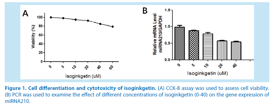
Vascular endothelial cells grew as a singlecell suspension. After treatment with 0-60 uM isoginkgetin for 36 h, we performed a CCK-8 assay to assess the cytotoxicity of isoginkgetin (Figure 1A). The effect of different concentrations of isoginkgetin (0- 40) on the gene expression of miRNA210 (Figure 1B).
Figure 1. Cell differentiation and cytotoxicity of isoginkgetin.
(A) CCK-8 assay was used to assess cell viability.
(B) PCR was used to examine the effect of different concentrations of isoginkgetin (0-40) on the gene expression of miRNA210.
Isoginkgetin significantly inhibits miRNA210 and PI3K/AKT signaling pathway in lipopolysaccharide induced human endothelial cells
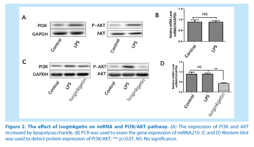
After 2 h of pretreatment with isoginkgetin 20 uM, human endothelial cells were treated with 1ug/LPS for 36 h. The results showed that lipopolysaccharide could increase the expression of PI3K and AKT (Figure 2A). However, Isoginkgetin had no significant effect on the expression of miRNA210 protein (Figure 2B). Isoginkgetin can significantly inhibit the gene expression of miRNA, besides, it can also significantly inhibit the PI3K/AKT signaling pathway in lipopolysaccharide induced human endothelial cells (Figure 2C and 2D).
**: p<0.01, NS: No significance.
Figure 2. The effect of isoginkgetin on miRNA and PI3K/AKT pathway.
(A) The expression of PI3K and AKT increased by lipopolysaccharide.
(B) PCR was used to exam the gene expression of miRNA210.
(C and D) Western blot was used to detect protein expression of PI3K/AKT.
Overexpression of miR-210 can significantly weaken the inhibitory effect of isoginkgetin on the PI3K/AKT signaling pathway in lipopolysaccharide induced human endothelial cells
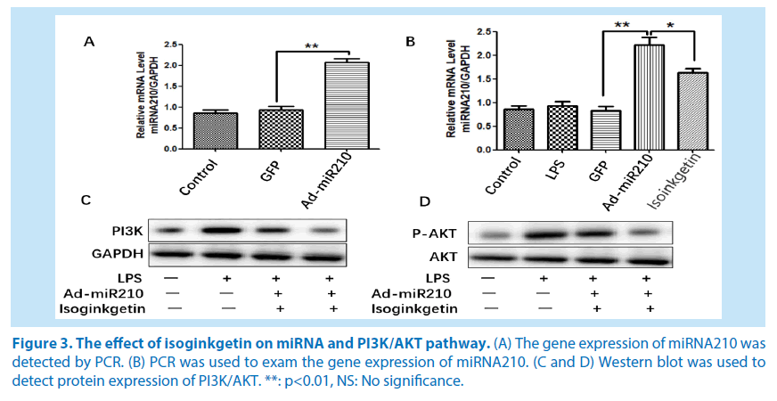
The recombinant adenovirus was transfected to HUVE-12 for 6 h when the cells proliferated to about 50%, and then the culture medium was replaced by isoginkgetin and lipopolysaccharide to treat endothelial cells. Green fluorescence was found under the fluorescence microscope, and the positive rate of green fluorescent protein (GFP) was above 50% after 36 h. The result showed that adenovirus can significantly overexpress the gene expression of miRNA210 (Figure 3A). The overexpressed miRNA can be significantly inhibited by isoginkgetin (Figure 3BD). After overexpression of miRNA in lipopolysaccharide treated endothelial cells, the effect of isoginkgetin on the inhibition of PI3K/AKT was significantly attenuated.
**: p<0.01, NS: No significance.
Figure 3. The effect of isoginkgetin on miRNA and PI3K/AKT pathway.
(A) The gene expression of miRNA210 was detected by PCR.
(B) PCR was used to exam the gene expression of miRNA210.
(C and D) Western blot was used to detect protein expression of PI3K/AKT.
Discussion
Ginkgo biloba is a rare plant in China. The isoginkgetin is an effective component extracted from the leaves of Ginkgo biloba. According to the literature, Ginkgo biloba leaves have the functions of dilating blood vessels, lowering serum cholesterol, and increasing the flow of coronary artery. Flavonoids extracted from Ginkgo biloba are natural antioxidants. Previous studies have proved that flavonoids can scavenge superoxide anion, hydroxyl radicals, lipid peroxidation free radicals and so on. Flavonoids can also slow down the formation of atherosclerosis and have an obvious antagonistic effect on the apoptosis of myocardial cells caused by ischemia [9].
With the in-depth study, it is found that miRNA plays an important role in cardiovascular events and cardiovascular function, which was first evidenced by Dicer gene knockout mice model: the fetus has hypoplastic heart and eventually died in embryonic period and Dicer knockout in heart of adult mouse leads to heart failure as well as death [10]. The finding suggests that miRNA plays an important role in the development of cardiovascular system and the maintenance of physiological function. Studies have found that [11-12] miRNA210 is hypoxia inducible miRNA. On the condition of hypoxia, the expression of miRNA210 is up-regulated in endothelial cells, then promotes the formation of capillary structure and cell migration while the inhibition of miR-210 can block angiogenesis. Studies have also shown that the expression of miR -210 is steadily up regulated after cerebral ischemia in the cortical area [13], which is up-regulated as well after renal ischemia in mice.
Recent researches have suggested that [14], PI3K/AKT signaling pathways are involved in both progression and angiogenesis of tumor. PI3K is a kind of intracellular signaling protein with catalytic activity. It can regulate the phosphorylation level of downstream AKT and the two together constitute PI3K/AKT signaling pathway, which plays a regulatory role in migration of a variety of cells [15]. Jiang et al. [16] found that overexpression of PI3K or AKT in chick embryos can induce angiogenesis and angiomegaly. However, it remains to be figured out whether miR -210 can regulate the PI3K/AKT signaling pathway.
In our present study, isoginkgetin can inhibit the gene expression of miRNA210, and inhibit the protein expression of PI3K/AKT signaling pathway in lipopolysaccharide induced endothelial cells; After overexpression of miRNA210, the role of isoginkgetin in PI3K/ AKT pathway was decreased significantly in lipopolysaccharide induced endothelial cells. The results showed that isoginkgetin mainly plays a role in inhibiting the PI3K/ AKT pathway through miRNA210. Our study provides experimental evidence for the study of the mechanisms of Ginkgo biloba in many diseases.
References
- Yoon SO, Shin S, Lee HJ et al. Isoginkgetin inhibits tumor cell invasion by regulating phosphatidylinositol 3-kinase/Akt-dependent matrix metalloproteinase-9 expression. Mol. Cancer. Ther. 5(11), 2666-2675 (2006).
- Sambandan S, Akbalik G, Kochen L et al. Activity-dependent spatially localized miRNA maturation in neuronal dendrites. Sci. 355(6325), 634-637 (2017).
- Ganju A, Khan S, Hafeez BB et al. miRNA nanotherapeutics for cancer. Drug. Disc. Today. 22(2), 424-432 (2017).
- Steudemann C, Bauersachs S, Weber K et al. Detection and comparison of microRNA expression in the serum of Doberman Pinschers with dilated cardiomyopathy and healthy controls. BMC. Vet. Res. 9(1), 12 (2013).
- Muralimanoharan S, Guo C, Myatt L et al. Sexual dimorphism in the effect of maternal adiposity on placental miR-210 expression. Placenta. 34(9), A38 (2013).
- Ono S, Oyama T, Lam S et al. A direct plasma assay of circulating microRNA-210 of hypoxia can identify early systemic metastasis recurrence in melanoma patients. Oncotarget. 6(9), 7053-7064 (2015).
- Giannakakis A, Sandaltzopoulos R, Greshock J et al. miR-210 links hypoxia with cell cycle regulation and is deleted in human epithelial ovarian cancer. Cancer. Biol. Ther. 7(2), 255-264 (2008).
- Ho A S, Huang X, Cao H et al. Circulating miR-210 as a novel hypoxia marker in pancreatic cancer. Trans. Oncol. 3(2), 109-113 (2010).
- Kwon CS, Kim JH, Son KH et al. Induction of cellular quinone reductase by some flavonoids. Food. Sci. Biotech. 12(6), 649-653 (2003).
- Cheng S, Zhang C, Xu C et al. Age-dependent neuron loss is associated with impaired adult neurogenesis in forebrain neuron-specific dicer conditional knockout mice. Int. J. Biochem. Cell. Biol. 57, 186-196 (2014).
- Xu TX, Zhao SZ, Dong M et al. Hypoxia responsive miR-210 promotes cell survival and autophagy of endometriotic cells in hypoxia. Eur. Rev. Med. Pharmacol. Sci. 20(3), 399-406 (2016).
- Muralimanoharan S, Guo C, Myatt L et al. Sexual dimorphism in miR-210 expression and mitochondrial dysfunction in the placenta with maternal obesity. Int. J. Obesity. 39(8), 1274-1281 (2015).
- Gan L, Liu Z, Wei M et al. MiR-210 and miR-155 as potential diagnostic markers for pre-eclampsia pregnancies. Med. 96(28), e7515 (2017).
- Xie Q, Wen H, Zhang Q et al. Inhibiting PI3K-AKt signaling pathway is involved in antitumor effects of ginsenoside Rg3 in lung cancer cell. Biomed. Pharmacother. 85, 16-21 (2017).
- Huang XL, Xu J, Zhang XH et al. PI3K/Akt signaling pathway is involved in the pathogenesis of ulcerative colitis. Inflammation. Res. 60(8), 727-734 (2011).
- Jiang BH, Zheng JZ, Aoki M et al. Phosphatidylinositol 3-kinase signaling mediates angiogenesis and expression of vascular endothelial growth factor in endothelial cells. Proc. Natl. Acad. Sci. USA. 97(4), 1749-1753 (2000).
