Case Report - Interventional Cardiology (2024)
Left atrial strain in the study of atrial extrasystole with delayed interatrial conduction: A Case report
- Corresponding Author:
- José Miguel Soler
Department of Cardiology, Nostra Senyora de Meritxell Hospital, Andorra laVella, Andorra,
E-mail: josepmiquel. soler@gmail.com
Received date: 22-Jul-2024, Manuscript No. FMIC-24-142824; Editor assigned: 24-Jul-2024, PreQC No. FMIC-24-142824 (PQ); Reviewed date: 08-Aug-2024, QC No. FMIC-24-142824; Revised date: 15-Aug-2024, Manuscript No. FMIC-24-142824 (R); Published date: 23-Aug-2024, ![]()
Abstract
We present the case of a 58-year-old woman CHADVASc 1 with very symptomatic palpitations in the form of frequent atrial extrasystole and short repetitive beats, initially studied with electrocardiography and echocardiography, including left atrial strain. The baseline electrocardiogram showed atrial extrasystole with probable origin around the pulmonary veins with increased duration of the P wave. The result of the echocardiographic study with segmental speckle tracking showed a sequence of images highly suggestive of an atrial mechanical block at the level of the Bachman´s bundle, which was confirmed by electroanatomical mapping (CARTO). This utility represents a novelty in the use of this technique in patients with atrial extrasystole in the absence of apparent organic heart disease.
Keywords
Left atrial strain • Atrial extrasystole • Bachmann’s bundle • Electrocardiogram • Interatrial block • Atrial fibrillation
Introduction
Although the description of the Bachman fascicle as a preferential electrical route in atrial conduction was made in 1916, it was not until the 1970s that it achieved greater clinical prominence in relation to its participation in the genesis and maintenance of certain atrial arrhythmias, performing new electrocardiographic and vectocardiographic mapping techniques [1], as well as experimental studies of endocavitary mapping in animals [2]. Subsequently, the obtaining of endocavitary recordings in humans showed preferential wavefronts in the cranio-caudal-right-left direction and their anomalies, reversing the depolarization of the left in a retrograde form from the lower area of the atrial septum when the blockage in the roof of the left atrium is advanced. Currently, the findings obtained with high-resolution mapping and postmortem examination [3], reaffirm the importance of this muscle band in the interatrial connection, being the most morphologically and electrophysiologically consistent pathway. The usefulness of echocardiography, and especially techniques with global longitudinal left atrial strain, has been suggested for clinical use in the most advanced cases of block [4], and the use of segmental strain remains largely unknown, although its usefulness has already been proposed in a study carried out by us previously [5].
Case Presentation
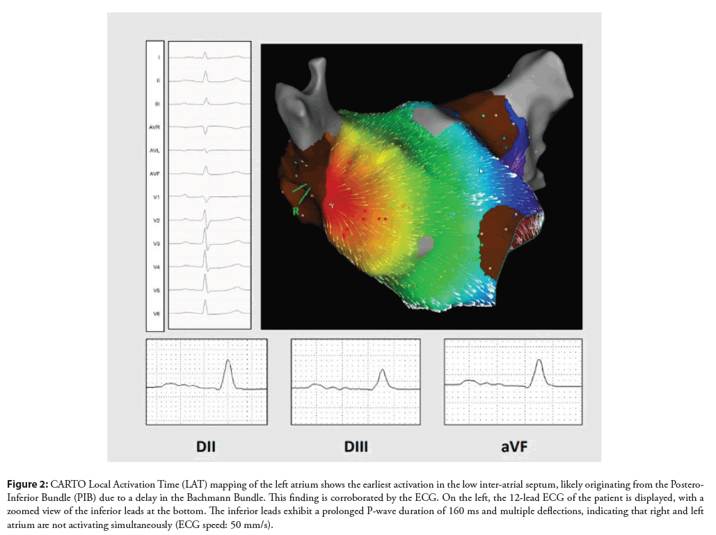
A 58-year-old female patient with a family history of ablated atrial fibrillation in two siblings and no other potentially premonitory family history. Occasional blood pressure elevations without diagnostic criteria for arterial hypertension in strict outpatient follow-up (hospital nurse) without requiring antihypertensive treatment (CHADVASc 1). First cardiological check-up was carried out in 2021 for very limiting short palpitations whose initial electrocardiograms showed a slight intermittent increase in the duration of the P wave with atrial extrasystole of probable origin in the vicinity of the pulmonary veins. 3-day electrocardiographic monitoring (χ2) recordings with high-density atrial extrasystole and frequent short repetitive forms of less than 3 seconds. The baseline echocardiographic study showed no alterations and the left atrial strain recordings incorporating an atrial extrasystole in several correlative frames pointed to a delay in electromechanical conduction at the level of the Bachmann’s bundle. She underwent a first electrophysiological study with a drop in atrial fibrillation during the beginning of the procedure, performing a first ablation of pulmonary veins. The persistence of the initial symptoms without changes in the continuous electrocardiographic monitoring records motivated a new ablation attempt in 2022 with RSV reconnection (roof, antero-superior and antero-inferior). The evolution was not entirely favorable, with short but limiting palpitations, maintaining frequent atrial extrasystole with short repetitive bursts, motivating a new extended electrophysiological study in 2024, where we found reconnection of the left (antero-superior) and right pulmonary vein(postero-inferior). We also carry out a CARTO Local Activation Time (LAT) map which confirmed the diagnosis done with Eco-TT as we found a delay in the Bachmann bundle with lower earliest activation of the left atrium. Re-ablation of the reconnection areas of the pulmonary veins as well as the fractional potentials in the left atrium was performed. Two months later, the patient has noticed an evident clinical improvement.
Results and Discussion
The relevant role of Bachmann’s bundle in maintaining atrial synchrony, its electrocardiographic description and its electrophysiological properties, as well as its involvement in the genesis of atrial arrhythmias, has been profusely documented [6-8]. Included in the concept of atrial dysfunction, it can develop areas of fibrosis even before atrial dilation, increasing the risk of atrial fibrillation at its maximum, and even posing its existence as an independent risk factor in thromboembolic pathogenesis. In the absence of atrial dilation, its occasionally intermittent appearance detracts from the diagnostic usefulness of the conventional electrocardiogram [9]. Long-term continuous electrocardiographic monitoring has been a very useful tool in the detection of arrhythmic complications at the outpatient level [10]. However, Dopplerechocardiography does not seem to have made major advances in the ambulatory study of atrial dysfunction in the early stages. Although the global and segmental basal left atrial strain has been shown to be useful in the detection of degrees of atrial myopathy prior to dilation [11], our experience has shown little value in cases with atrial extrasystole and/or short repetitive forms in the absence of atrial fibrillation [5].
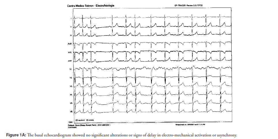
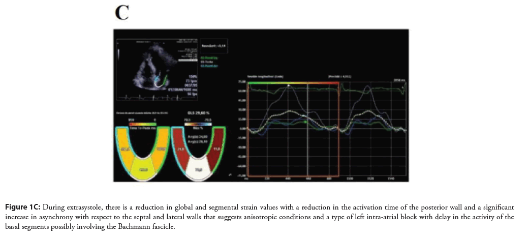
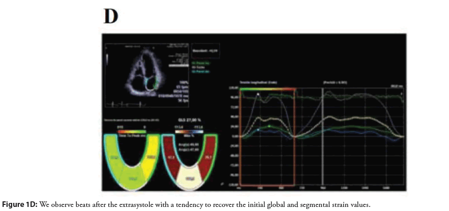
The possibility of synchronizing the presence of an atrial extrasystole with the serial frames of echocardiography and left atrial strain is, in our opinion, a possible major advance in the detection of initial degrees of left atrial dysfunction and alterations in interintraatrial conduction in particular (Figures 1A-1D). The basal electrocardiogram showed small alterations in the durability of the P wave and the extrasystole was positive on the lower side and V1 with negativity in aVL suggestive of an origin in the posterior left superior pulmonary vein.
Figure 2: Shows the deformation curves of the septal, posterior and lateral walls during sinus rhythm, showing a maximum peak of positive longitudinal elongation. Very predominant posterior aspect with slight asynchrony with respect to the activation of the septal and lateral walls (18 ms) with reservoir strain values within normal ranges.
Figure 3: During extrasystole, there is a reduction in global and segmental strain values with a reduction in the activation time of the posterior wall and a significant increase in asynchrony with respect to the septal and lateral walls that suggests anisotropic conditions and a type of left intra-atrial block with delay in the activity of the basal segments possibly involving the Bachmann fascicle.
Parietal stress, intracavitary pressure changes, and changes in electromechanical activation sequences, independent of left ventricular function, can help detect subtle abnormalities in conduction not detectable by other noninvasive techniques. Its study will allow a better understanding of the mechanisms of left atrial adaptation and a better selection of patients who are candidates for electrophysiological study and ablation techniques (Figures 2 and 3).
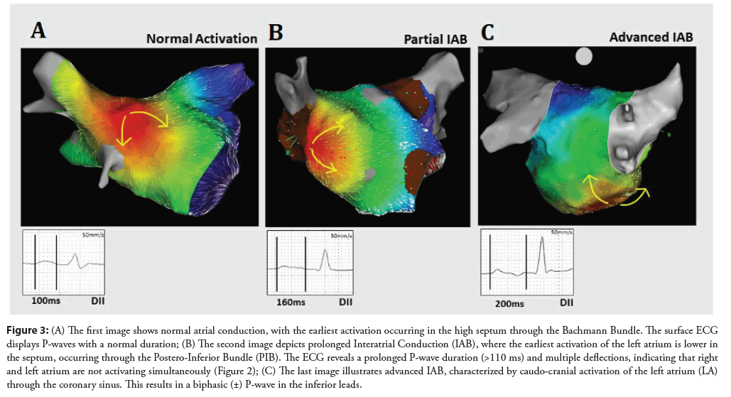
Figure 2: CARTO Local Activation Time (LAT) mapping of the left atrium shows the earliest activation in the low inter-atrial septum, likely originating from the Postero- Inferior Bundle (PIB) due to a delay in the Bachmann Bundle. This finding is corroborated by the ECG. On the left, the 12-lead ECG of the patient is displayed, with a zoomed view of the inferior leads at the bottom. The inferior leads exhibit a prolonged P-wave duration of 160 ms and multiple deflections, indicating that right and left atrium are not activating simultaneously (ECG speed: 50 mm/s).
Figure 3: (A) The first image shows normal atrial conduction, with the earliest activation occurring in the high septum through the Bachmann Bundle. The surface ECG displays P-waves with a normal duration; (B) The second image depicts prolonged Interatrial Conduction (IAB), where the earliest activation of the left atrium is lower in the septum, occurring through the Postero-Inferior Bundle (PIB). The ECG reveals a prolonged P-wave duration (>110 ms) and multiple deflections, indicating that right and left atrium are not activating simultaneously (Figure 2); (C) The last image illustrates advanced IAB, characterized by caudo-cranial activation of the left atrium (LA) through the coronary sinus. This results in a biphasic (±) P-wave in the inferior leads.
The expert consensus on atrial cardiomyopathy proposed the following as a working definition of this: “any complex of structural, architectural, contractile, or electrophisological changes affecting the atria with the potential to produce clinicallyrelevant manifestations”. It could, therefore, be considered that the contractile alterations of the left atrium and the occurrence of supraventricular arrhythmias in patients with interatrial block would be a form of atrial cardiomyopathy [12].
Conclusion
We present a variant in the study of echocardiography with left atrial strain in a selected patient with delayed conduction at the level of the Bachmann fascicle. The inclusion of an atrial extrasystole and the sequence integrated with the multi-frame electrocardiogram can be very useful in diagnostic and prognostic stratification in outpatients without structural heart disease.
References
- Castillo.A, Vernant.P. Troubles de la conduction intrauricular par bloc du fasceau de Bachmann. Arch Mal Coeur Vaiss.64:1490-1503 (1971).
- Allessie MA, Bonke FI, Schopman FJ, et al. Circus movement in rabbit atrial muscle as a mechanism of tachycardia. III. The" leading circle" concept: A new model of circus movement in cardiac tissue without the involvement of an anatomical obstacle. Circ Res.41(1):9-18 (1977).
- Knol WG, Teuwen CP, Kleinrensink GJ, et al. The bachmann bundle and interatrial conduction: Comparing atrial morphology to electrical activity. Heart Rhythm.16(4):606-614 (2019).
- Teuwen CP, Yaksh A, Lanters EA, et al. Relevance of conduction disorders in Bachmann’s bundle during sinus rhythm in humans. Circ Arrhythm Electrophysiol.9(5):e003972 (2016).
- Soler JM, García-Parés G, Valero O, et al. Assessment of short forms of recurrent atrial extra systoles by echocardiography with left atrial strain in ambulatory patients without organic cardiopathy. Arch Cardiol Mex. 93(2):172-182 (2023).
- Bejarano-Arosemena R, Martínez-Sellés M. Interatrial block, Bayés syndrome, left atrial enlargement, and atrial failure. J Clin Med.12(23):7331 (2023).
- Chhabra L, Devadoss RJ, K Chaubey VA, et al. Interatrial block in the modern era. Curr Cardiol Rev.10(3):181-189 (2014).
- Ariyarajah V, Fernandes J, Kranis M, et al. Prospective evaluation of atrial tachyarrhythmias in patients with interatrial block. Int J Cardiol.118(3):332-337 (2007).
- Bayés de Luna A, Baranchuk A, Niño Pulido C, et al. Secondâdegree interatrial block: Brief review and concept. Ann Noninvasive Electrocardiol.23(6):e12583 (2018).
- Zepeda-Echavarria A, van de Leur RR, van Sleuwen M, et al. Electrocardiogram devices for home use: Technological and clinical scoping review. JMIR cardio.7:e44003 (2023).
- O’Neill T, Kang P, Hagendorff A, et al. The clinical applications of left atrial strain: A comprehensive review. Medicina.60(5):693 (2024).
- Goette A, Kalman JM, Aguinaga L, et al. EHRA/HRS/APHRS/SOLAECE expert consensus on atrial cardiomyopathies: Definition, characterization, and clinical implication. Ep Europace.18(10):1455-1490 (2016).
