Clinical images - International Journal of Clinical Skills (2019) Volume 13, Issue 1
Localized Nodular Adiposis Dolorosa in Patient with Rheumatoid Arthritis
- Corresponding Author:
- Karla Asturias
Universidad Francisco Marroquín School of Medicine, Guatemala City, Guatemala
E-mail: kasturias@ufm.edu
Abstract
A 58-year-old female with no previous medical history presented with a 10-year history of recurrent bilateral metacarpophalangeal and proximal interphalangeal joint pain and inflammation, associated with morning stiffness.
Keywords
Clinical examination; Medical; Pain; Inflammation
Clinical-Medical Examination
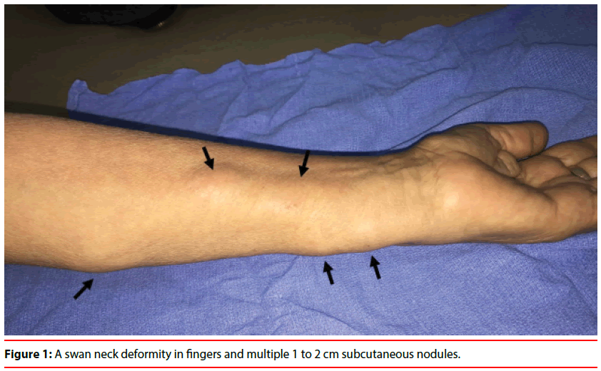
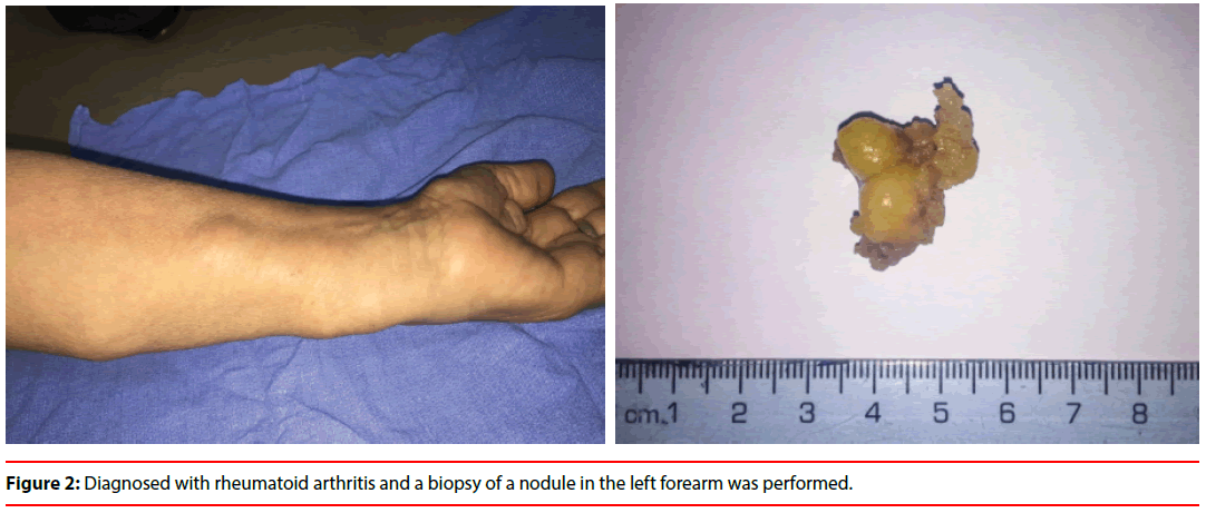
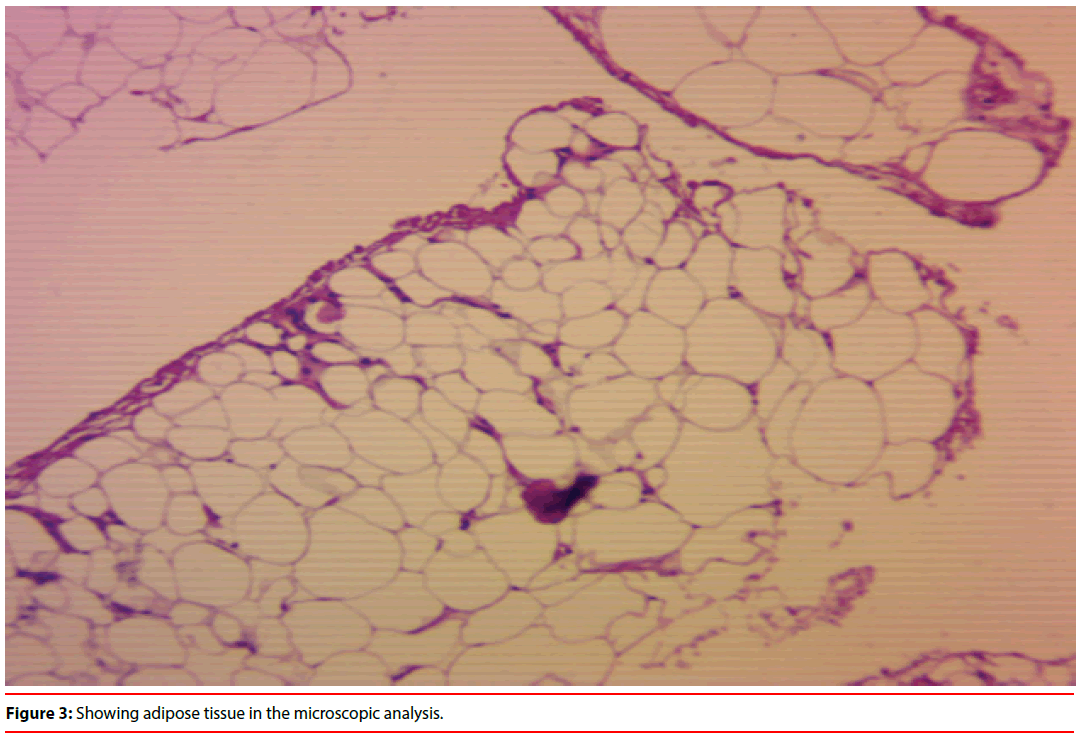
A 58-year-old female with no previous medical history presented with a 10-year history of recurrent bilateral metacarpophalangeal and proximal interphalangeal joint pain and inflammation, associated with morning stiffness. Early in the course of her disease, she presented to a primary care clinic but did not followed-up due to economic reasons. At the time of this presentation, physical examination showed ulnar deviation bilaterally, a swan neck deformity in fingers and multiple 1 to 2 cm subcutaneous nodules (Figure 1) affecting the forearms, that were soft, tender and moveable, distributed bilaterally in a non-symmetric fashion, with no associated skin changes. Nodules elicited discomfort and dull pain in the affected areas. Laboratory testing was remarkable for a rheumatoid factor <8 IU/mL and anti-cyclic citrullinated peptide of 1033 U/mL. She was diagnosed with rheumatoid arthritis and a biopsy of a nodule in the left forearm was performed (Figure 2), showing adipose tissue in the microscopic analysis (Figure 3). Alternative diagnoses were excluded, and adiposis dolorosa or Dercum’s disease, with its localized nodular form, was considered as the main differential diagnosis. She was treated symptomatically, and her rheumatoid arthritis regimen was recently started.
Figure 2: Diagnosed with rheumatoid arthritis and a biopsy of a nodule in the left forearm was performed.
Figure 3: Showing adipose tissue in the microscopic analysis.


