Research Article - Imaging in Medicine (2020) Volume 12, Issue 5
Magnetic Resonance Imaging Nomogram For Optic Nerve And Extraocular Muscles In Nigerian Subjects
Valentine C Ikamaise1, Amabe O Akpantah2, Aniekan A Jacob3, Bassey E Archibong1* & Eusebius Dike U41Department of Radiography & Radiological Science, University of Calabar, Nigeria
2Department of Anatomical Sciences, University of Calabar, Nigeria
3Department of Radiology, University of Uyo Teaching Hospital, Akwa Ibom State, Nigeria
4Department of Radiology, National Hospital, Abuja, Nigeria
- Corresponding Author:
- Bassey E Archibong
Department of Radiography & Radiological Science
University of Calabar, Nigeria
E-mail: bassey.archibong@unical.edu.ng
Abstract
Background: The role of optic nerves and extraocular muscles as critical in conveying visual signals to the brain and controlling movement of the eyeballs to ensure precise vision cannot be overemphasized.
Objective: The study aimed at the assessment of optic nerves and extraocular muscles of 195 brain images generated by Magnetic Resonance Imaging (MRI) to establish normal sizes of these tissues.
Methods: Direct measurement of the size of optic nerves and extraocular muscles was carried out respectively on the magnetic resonance images with the aid of a Digital Imaging and Communications in Medicine (DICOM) image viewer. The data was analyzed using Microsoft Excel 2007 by subjecting to descriptive statistics, student’s t-test and Pearson’s correlation statistical tools.
Result: The mean size of the right optic nerve of the subjects was 4.89 ± .77mm and the left was 4.87 ± .69mm. There was no significant difference (p>0.001) between the size of the right and the left optic nerves. The sizes of extraocular muscles of subjects studied were: 3.15 ± .49mm for Right Lateral Rectus Muscle (RLRM); 3.06 ± .46mm for Left Lateral Rectus Muscle (LLRM); 3.13 ± .39mm for Right Medial Rectus Muscle (RMRM) and 3.20 ± .39mm for Left Medial Rectus Muscle (LMRM). There were no significant differences between sizes of extraocular muscles (p<0.001).
Conclusion: The findings have provided a standard for future quantitative comparison and will serve as predictive indicators or radiologic reference for the diagnosis of patient with associated visual problems.
Keywords
Extraocular muscles ■ Medial rectus muscle ■ Optic nerve ■ Extraocular muscles
Introduction
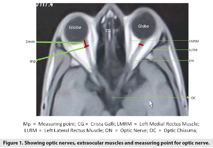
The functional units of the visual pathway include the optic nerve which is a paired nerve that conveys visual impulse from the retina to the brain. The optic nerve begins at the disc, behind the retina and runs posteriorly to the optic chiasma. (FIGURE 1). Beyond the optic chiasma the optic nerve continues as the optic tract to the visual cortex in the brain [1]. The fibers of the optic nerve are a posterior continuation of ganglion nerve fiber layer of the retina that specializes in sensing light and signaling the optic nerve fibers. Each of the optic nerves contains about 1.2 million fibers [2,3].
The optic nerve is visualized as a hypo-intense (light grey) stripe on an MR generated image running through the length of the orbit behind the eyeball from the disc postero-inferiorly to the optic chiasma (FIGURE 1). The nerve is clearly outlined on both sides by the brighter density band of the optic nerve sheath covering the nerve through its length [4]. Significant variations in optic nerve morphology in homogenous population as well as race was reported by Selhorst and Chen [1]. In any event of damage to the optic nerve due to trauma, infection or congenital anomalies, loss of sight will ultimately occur due to its impact on the morphology of the optic nerve [5-13]. The above account underpins the need to determine the normal values and morphology of the optic nerves for the sake of the diagnostic ideals involved.
The extraocular muscles are six small, strong, and prearranged structures around the eyeball [14]. The extraocular muscles are specifically made up of the fast-twitch muscle fibrils which can turn the eye in various directions. Their action moves the eye backward and forward, sideways, up, and down, and around [15]. Extraocular muscles are superior rectus, superior oblique, inferior rectus, inferior oblique, lateral rectus, and medial rectus muscle.
The largest of these muscles is the medial rectus that coordinates the movements of the eyeball. The size is probably due to the frequency of use for convergence of the eye [16,17]. In moving the eye up and down to keep the pupil closer to the midline of the body, side to side and, in and out the muscles work in synergy to accomplish these tasks. Strabismus (Crossed eye) is the misalignment of the eye that results from malfunctioned medial rectus.
The lateral rectus that controls the movement of the pupil of the eye away from the midline of the body is inserted into the outer side of the temporal aspect of the eyeball and extends to the annulus of Zinn [18]. Studies on the determination of diameter and width of the muscles tendon have shown that Lateral rectus muscle (LRM) can be used as a source to generate information on diagnosis and interventions in cases like exotropia. Exotropia is an abnormal turning outward of one or both eyes balls. Trauma to LRM is rare. However, when it occurs it leads to exotropia even after successful management of the injuries [19,20]. Similarly, information on the morphologic and the clinical status of these muscles can be used to diagnose problems leading to vision loss.
All medical imaging techniques have tremendous contributions in optic nerve and extraocular muscles evaluation, but the advent of MRI as a new diagnostic tool in optic nerve neuropathy has opened up new grounds in unbiased and sophisticated evaluation of optic nerve structure in health and in sickness [21,22]. The advantage of natural soft tissue contrast enhancement of MRI over other imaging techniques has made MRI exceptional in investigative research involving optic nerve [23,24]. It enhances assessment of features of the optic nerve necessary for accurate diagnosis and monitoring of optic nerve associated neuropathy. This study, therefore, seizes the opportunity to explore the advantages inherent in MRI for a noninvasive direct measurement of optic nerve and extraocular muscles to determine the diameter as well as the width of these tissues and analyze the morphological relationship between them.
Measurements carried out with MRI on optic nerve and other tissues are used to establish normal sizes and variations in sizes for Caucasians, Asians, American and Afro- American [25]. Data on Africans and specifically on Nigerians are known to be scarce. The use of MRI-brain images in Nigerian hospitals to determine the size of optic nerve and extraocular muscles on subjects is necessary and timely due to its medical importance to Nigeria. This will in addition provide indigenous data to the study of optic nerve and extraocular muscles in the pursuit of the needed local content in research.
Methods
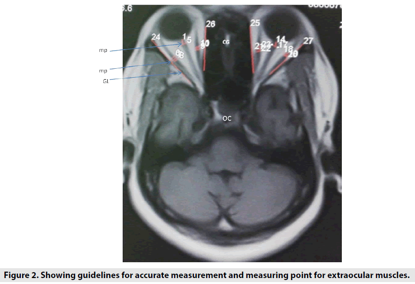
The measurement of the size of optic nerve and extraocular muscles was carried out retrospectively on 195 MRI brain images from Radiology Departments of a tertiary health facility in Nigeria. The sample size was based on application of Yaro Yamane formula for finite population sample size determination on a total number of normal brain scans for 2015 and 2016 [26], The hospital was selected based on availability of a functional MRI unit at the time of the study, and its status as a referral centre for health facilities within the Federal Capital territory, the North Central Geopolitical zone and the entire Northern Nigeria. A Phillips NT10 MRI scanner (0.3T) capable of generating high diagnostically acceptable images was used for the measurement. MRI brain images were retrieved from the facility storage vault and uploaded on a personal computer from where measurement was carried out. Techniques for the measurement of optic nerve is consistent and was carried out at a defined location to determine the size of optic nerve and extraocular muscles [27-29]. Viewing, manipulation and measurement on MRI images was done using K-PACS image viewer (2017 version). The image series of a subject was retrieved and displayed on a computer screen. The slice that presented the best image for optic nerve, lateral rectus muscle and medial rectus muscle was selected by flipping through a series of axial scanned images of the brain. Among the images in the T1 weighted series, the slice that displayed the image of intracranial section of the optic nerve that extends from the globe and enters medially into the optic canal located in the lesser wing of the sphenoid bone in profile was selected. Eyeballs were demonstrated with equal distance on both sides from crista galli on the selected slice. The optic nerve was visualized through the entire length from the optic foramen to the optic disk and the named extraocular muscles were also visualized through its length (FIGURE 2).
A point 3mm posterior to the optic disc or optic nerve head was located and marked. (FIGURES 1 & 2) [29]. The electronic caliper was placed at 90 degrees to the long axis of the optic nerve to accurately measure the diameter of the optic nerve. The placement of the calliper accurately on the image of optic nerve was facilitated by the guideline drawn parallel to the long axis of the nerve (FIGURE 2). Both optic nerves were measured repeatedly. An average of 3 measurements for each nerve and extraocular muscle was calculated and recorded to minimize observer’s error and ensure reproducibility of the measurement. The images generated on axial plane were chosen because it is associated with the least inter- and intra-observer variability [29]. Demographic and clinical information were obtained from the clinical records of the patients. Radiologist’s report on each case was examined to rule out the inclusion of the subjects with optic nerve and extraocular muscles pathology. All images that came out with positive outcome were excluded from data used to establish normal values.
The widths of the lateral and medial rectus muscles were measured on the same slice of the axial T1 weighted MRI brain image that the optic nerve measurement was taken. The selection and display of image after scans for measurement of the lateral and medial recti muscles was like that for optic nerve. Taking measurement of the width of extraocular muscles in the axis perpendicular to the orbital wall is the standard practice globally for computed tomography and MRI generated images. [27, 28]. In keeping with the global benchmark, measurement was carried out at where the muscle has its maximum diameter (this in most cases correspond to the midpoint on the length of the muscle). Practically, the calliper is used to draw a line parallel to an imaginary line passing through the mid plane of the muscle through its length. This line is called the “guide” and is used to direct the line that measures the diameter to be perpendicular to the length of the measured muscle (FIGURE 2). Each measurement generally was repeated three times. In the end, 18 measurements were made on one image amounting to a total of 3510 measurements from which means of each set of three measurements were computed and used for analysis.
Statistics tools for determination of central tendency and range were applied on the data to determine normal values of the tissues under study. Student t-test was applied on two sets of measurements to determine the differences. Comparability, association, and relationship were tested by Pearson’s product moment correlation coefficient.
Discussion
This study carried out a total of 3510 measurements of the diameter of optic nerves and the width of named extraocular muscles (medial and lateral recti muscles) on MRI generated brain images of 195 subjects. Normal values for optic nerves diameter and width of medial and lateral recti muscles were determined for subjects studied in Abuja (TABLE 1). These values are documented as reference values for diagnostic and treatment purposes. The measurement carried out in this study was at reference point of 3mm posterior to the optic disc or head, which is the standard reference point for determination of the normal diameter of the optic nerve by Computed Tomography (CT), ultrasound and MRI [29]. The values obtained in this study are not expected to be the same with reported values in literature because other researchers carried out measurements at various points on the track of the nerve and the choice of the measurement point depended on their objectives. Lagreze et al., reported 3.11 ± 0.33mm at 5mm point from the optic disc, 2.66 ± 0.30mm at 10mm point and 2.64 ± 0.24mm at 15mm point from the optic disc [30]. Their values are less than what is obtained in this study (5.14 ± 0.79mm and 5.23 ± 0.88mm for right and left respectively) because 3mm on the track of the nerve is where the nerve is largest in diameter.
| Tissue | Mean±SD (mm) | Range (mm) | Right & Left | Relationship Right medial & Lateral | Left medial & lateral | Significance |
|---|---|---|---|---|---|---|
| RON | 4.89 ± .77 | 2.80-7.53 | ||||
| LON | 4.87 ± .69 | 3.00-7.30 | r=.840** | P<0.001 | ||
| RLRM | 3.15 ± .49 | 2.20-4.93 | ||||
| RMRM | 3.13 ± .46 | 2.03-5.50 | r=.677** | P<0.001 | ||
| LLRM | 3.06 ± .46 | 2.23-4.37 | ||||
| LMRM | 3.20 ± .39 | 2.17-4.70 | r=.573** | P<0.001 | ||
| RLRM | 3.15 ± .49 | 2.20-4.93 | ||||
| LLRM | 3.06 ± 46 | 2.23-4.37 | r=.573** | P<0.001 | ||
| RMRM | 3.13 ± .46 | 2.03-5.50 | ||||
| LMRM | 3.20 ± .39 | 2.17-4.70 | r=.649** | P<0.001 | ||
| ** = Positive and strong correlation RON = Right optic nerve LON = Left optic nerve RLRM = Right left rectus muscle RMRM = Right medial rectus muscle LLRM = Left lateral rectus muscle RLRM = Right lateral rectus muscle |
||||||
Table 1. Normal sizes and relationship of optic nerves and named extraocular muscles for subjects studied (n = 195).
The diameter of both the left and right optic nerves of the subjects studied are not significantly different (p<0.001). Similarly, the width of the medial and lateral recti muscle measured on the same subject were not significantly different (p<0.001). One may not expect differences in the measured structure because the growth and development of measured tissues are identical since they exist as paired tissues. Other researchers may not have categorically compared the variations between sizes of left and right optic nerves and sizes of left and right extraocular muscles, but they have published dimensions on sizes of various parts of the optic nerves and extraocular muscles. Ogbole et al [31] reported the following diameters for extraocular muscles by a CT measurement in a population of normal patients in a tertiary hospital in X:
Medial Rectus = 4.5 ± 1.2 mm
Lateral Rectus = 4.9 ± 2.1 mm
Inferior Rectus = 4.8 ± 1.6 mm
Superior group = 4.0 ± 1.5 mm
The diameters of the medial and lateral recti muscles reported by Ogbole et al. [31] are larger than 3.52 ± 0.61 mm and 3.37 ± 0.55 for medial and lateral recti muscles respectively obtained by this study. This may probably be due to variations observed when different modalities are used to measure the same structure. Establishment of normal values is importance as they serve as radiological reference values in the assessment of changes in size. Measurements of optic nerve diameter (OND) and optic nerve sheath diameter (ONSD) are reported to be useful predictive indicators in occurrence of high pressure in the cranium, leading to raised intracranial pressure (ICP) and intracranial hypertension [32].
The implication of the strong and positive correlation of normal values of optic nerves diameter and width of extraocular muscles of either side recorded in this study show the synergy that exist in action between these structures (TABLE 1). This means that the loss or gain in action (status) of one can predict the status of the other. In the case of the optic nerves one will expect that if one of the nerves is absent, the other will acquire morphological strength to cope with increase demand. Incidentally, it was observed that in few subjects with only one optic nerve the size was less than normal. This may be understood or explained by application of programmed cell dead theory (apoptosis) [33].
A marked variation in the size of diameter and width of optic nerves, as well as extraocular muscles respectively was not observed within subjects.
Other researchers have reported on works aimed at correlation of optic nerve head area, axons in the optic nerve head and Retinal Nerve Fibre Layer (RNFL) with other related tissues. Frisen and Quigley reported some levels of relationship of Visual Acuity (VA) to axons that survive in optic nerves after an insult [34]. Their finding made it possible to estimate the functional fraction of foveo cortical neural channel from clinical acuity measurement. Nobuko [35] reported correlative relationship between the optic nerve head area and macular thickness (ɼ =0.225), RNFL thickness (ɼ = 0.253) and visual field defects in patients with Primary Open Angle Glaucoma (POAG) by spectral domain Optical Coherence Tomography (OCT). Their work revealed that Optic Nerve Head (ONH) significantly and positively correlated with macular and retinal nerve fibre layer. This means that the size of optic nerve head can be used to explain the thickness of both, the macular and RNFL in patients with primary open angle glaucoma.
Results
The diameter of the right optic nerve ranged between 2.80 mm – 7.53 mm with a mean value of 4.89 ± .77mm and the left optic nerves ranged between 3.00 mm – 7.30 mm with a mean of 4.87 ± .69 mm. (TABLE 1). The diameters of the right and the left optic nerves correlated strongly and positive with ɼ = 0.840 (P<0.01) in their relationship (TABLE 1). No significant different between the diameters of the right optic nerves (mean value of 4.89 ± .77 mm) and the left optic nerve (means value of 87 ± .69 mm) at P<0.001 within subjects studied. (TABLE 2).
| Tissue | Mean±SD (mm) | t-Value | P-Value | Significance |
|---|---|---|---|---|
| RLRM | 3.14 ± .49 | 0.359 | 0.72 | P<0.001# |
| RMRM | 3.13 ± .46 | |||
| LLRM | 3.06 ± .46 | -4.846 | 0 | P<0.001# |
| LMRM | 3.20 ± .39 | |||
| RLRM | 3.14 ± .49 | 2.661 | 0.008 | P<0.001* |
| LLRM | 3.06 ± 46 | |||
| RMRM | 3.13 ± .46 | -2.489 | 0.014 | P<0.001# |
| LMRM | 3.20 ± .39 | |||
| * = Significant # = Not significant RON = Right optic nerve LON = Left optic nerve RLRM = Right left rectus muscle RMRM = Right medial rectus muscle LLRM = Left lateral rectus muscle RLRM = Right lateral rectus muscle |
||||
Table 2. Comparison of optic nerve diameter and the width of named extraocular muscles within subjects studied.
The width of the right lateral rectus muscle ranged between 2.20 mm - 4.93 mm and a mean value of 3.15 ± .49 mm; the width of the right medial rectus muscle ranged between 2.30 mm – 5.50 mm with a mean of 3.13 ± .46 mm; for the left lateral rectus muscles the range of 2.23mm – 4.37mm and a mean of 3.06 ± .46mm was obtained. The width of the left medial rectus muscle ranged between 2.17mm – 4.70 mm and a mean value of 3.20 ± .39 mm. (TABLE 1). The width of these extraocular muscles showed strong and positive relationship among themselves with ɼ = not less than 0.573 (p<0.01) for subjects studied (TABLE 1).
The width of the lateral and medial rectus muscles (3.15 ± .49 mm) and (3.13 ± .46 mm for right lateral rectus muscle and right medial rectus muscle; 3.06 ± .46 mm and 3.20 ± .39 mm for left lateral rectus muscle and left medial rectus muscle were not markedly different within subjects studied at p<0.001 (TABLE 2).
Conclusion
This study aimed at measuring the optic nerves and extraocular muscles to determine their sizes in diameter and width respectively on MRI generated images in a health facility at Abuja, Nigeriay. After a successful measurement and statistical analysis carried out on 195 subjects the following conclusion can be drawn. The normal size in diameter for right optic nerve was 5.14 ± 0.79 mm and left optic nerve was 5.23 ± 0.88 mm. there was no marked variations in size between left and right optic nerves. The normal size in width for right lateral rectus muscle was 3.37 ± 0.55 mm, right medial rectus muscle was 3.52 ± 0.61 mm, left lateral rectus muscle was 3.58 ± 0.68 mm and left medial rectus muscle was 3.67 ± 0.88 mm. The study has provided a nomogram for future comparison of optic nerve and extraocular muscles measurements. These will serve as predictive indicators or radiologic reference for the diagnosis of patients with visual problems associated with studied parameters.
Acknowledgements
None
Funding
This research received no specific grant from any funding agency in the public, commercial, or not-for-profit sectors.
Declaration of interests
None.
References
- Selhorst JB, Chen Y. The optic nerve. Semin. Neurol. 29, 29-35, (2009).
- Jonas JB, Schmidt AM, Muller-Bergh JA et al. Human optic nerve fiber count and optic disc size. Investig. Ophthalmol. Vis. Sci. 33, 2012-8, (1992).
- Balazsi AG, Rootman J, Drance SM et al. The effect of age on the nerve fiber population of the human optic nerve. Am. J. Ophthalmol. 97, 760-766, (1984).
- Shevlin C. Optic Nerve Sheath Ultrasound for the Bedside Diagnosis of Intracranial Hypertension: Pitfalls and Potential. Crit. Care. Hor. 1, 22-30, (2015).
- Larson B. Visual fields defects secondary to a cerebrovascular accident. Opto. Vis. Per. 3, 307-316, (2015).
- Esther MH, Linda MZ, Johnathan G et al. Optic disk size & glaucoma. Surv. Opthalmol. 52, 32-49, (2006).
- Robin L. Gal MSPH. “Multiple Sclerosis risk after optic neuritis: Final optic neurirtis treatment trial follow-up”. Arch. Neurol. 65, 727-32, (2008).
- Catherine U. Ukponmwan. Pattern of ocular morbidity in Nigeria. Asian. Pac J. Trop. 3, 164-166, (2013).
- Ogun OA, Adediran OA. Non-glaucomatous optic neuropathy in Ibadan: extrapolations to healthcare funding in Nigeria. Ann. Ib. Postgrad. Med. 12, 103-108, (2014).
- Amador-Patarroyo MJ, Perez-Rueda MA, Tellez CH. Congenital anomalies of the optic nerve. Saudi. J. Ophthalmol. 29, 32-38, (2015).
- https://emedicine.medscape.com/article/1217005-clinical
- Osaguona V, Okeigbemen V. Non-glaucomatous optic atrophy in Benin City. Ann. Afr. Med. 14, 109-113, (2015).
- Boland MV, Lee IH, Zan E et al. Quantitative Analysis of the Displacement of the Anterior Visual Pathway by Pituitary Lesions and the Associated Visual Field Loss. Investig. Ophthalmol. Vis. Sci. 57, 3576-3580, (2016).
- Ridyard E. Extraocular Muscles: Variation in Their Anatomy, Length and Cross-Sectional Diameter. Int. J. Anat. Res. 3, 1198-1206, (2015).
- Hao R, Suh SY, Le A et al. Rectus Extraocular Muscle Size and Pulley Location in Concomitant and Pattern Exotropia. Ophthalm. 123, 2004-2012, (2016).
- Duranoglu Y, Ilhan HD, Guler AM. Surgical results of the slipped medial rectus muscle after hanging back recession surgery. Int. J. Ophthalm. 7, 1035-1038, (2014).
- Toole LO, Long V, Power W et al. Traumatic rupture of the lateral rectus. Eye. 18, 221-222, (2004).
- Yun CM, Kim SH. The tendon width of lateral rectus muscle in predicting the effect of recession: Is it just age-related artefact. Eye. 25, 1356-1359, (2011).
- Newman WD, Hollman AS, Dutton GN et al. Measurement of optic sheath diameter by ultrasound: a means of detecting acute raise intracranial pressure in hydrocephalus. Brit. J. Ophthalmol. 86, 1109-1113, (2002).
- Benjamin JO, Nicholas JV. Optic neuritis and risk of MS: differential diagnosis and management. Cleve. Clin. J. Med. 76, 181-90, (2009).
- Costello F, Hodge W, Irene Pan Y. Exploring the association between retinal nerve fiber layer thickness and initial magnetic resonance imaging findings in patients with acute optic neuritis. Mult. Scler. Int. (2011).
- Samarawickrama C, Hong T, Jonas JB et al. Measurement of Normal Optic Nerve Head Parameters. Surv. Ophthalmol. 57, 317-336, (2012).
- Sobol WT. Recent advances in MRI technology: Implications for image quality and patient safety. Saudi. J. Ophthalmol. 26, 393-399, (2012).
- Etman HM, Mokhtar A, Abd-Elhamid MI et al. The effect of quality control on the function of magnetic resonance imaging (MRI), using American College of Radiology (ACR) phantom. Egy. J. Radiol. Nucl. Med. 48, 153-160, (2017).
- Christopher AG, Gerald MMS, Michael JS et al. Variation in optic nerve and macular structure with age and race with spectral-domain optical coherence tomography. Ophthalmol. 118, 2403-2408, (2011).
- Uzoagulu AE, Yaro. Yamane formula for a finite population. Pract. Guide. Writ. Res. Proj. Rep. Tertiary. Inst. 57-59, (2011).
- Ozgen A, Aydingoz U. Normative measurements of orbital structures using MRI. J. Comp. Ass. Tomograp. 24, 493-496, (2000).
- Lee JP, Park IW, Chung YS. The volume of tumour mass and visual field defect in patients with pituitary macroadenoma. Korean. J. Ophthalmo. 25, 37-41, (2011).
- Bauerle J, Schuchardt F, Schroeder L et al. Reproducibility and accuracy of optic nerve sheath diameter assessment using ultrasound compared to magnetic resonance imaging. BMC. Neurol. 13, 187, (2013).
- Lagreze W. Treatment of optic neuropathies-state of the art. Klin. Monatsbl. Augenh. 226, 875-880, (2009).
- Ogbole GI, Ogun OA, Olusumade D. Computed tomography measurement of extra-ocular muscle diameters in a population of normal patients in a tertiary hospital in Nigeria. Afr. J. Med. Sci. 43, 245-250, (2014).
- Geeraerts T, Newcombe VFJ, Coles JP et al. Use of T2-weighted magnetic resonance imaging of the optic nerve sheath to detect raised intracranial pressure. Crit. care. 12, 114, (2008).
- Shofty B, Ben-Sira L, Constantini S et al. Optic nerve sheath diameter on MR imaging: establishment of norms and comparison of pediatric patients with idiopathic intracranial hypertension with healthy controls. Am. J. Neuroradiol. 33, 366-369, (2012).
- Frisén L, Quigley HA. Visual acuity in optic atrophy: a quantitative clinicopathological analysis. Graef. Arch. Clin. Exp. Ophthalmol. 222, 71-74, (1984).
- Enomoto N, Anraku A, Ishida K et al. Size of the Optic Nerve Head and Its Relationship with the Thickness of the Macular Ganglion Cell Complex and Peripapillary Retinal Nerve Fibre Layer in Patients with Primary Open Angle Glaucoma. J. Ophthalmol. 15, 186-249, (2015).


