News and Views - Imaging in Medicine (2011) Volume 3, Issue 1
Magnetic resonance spectroscopy provides possible diagnosis of brain disorders suffered by athletes
Emily Thornton*University of California, San Francisco, CA, USA
Abstract
Magnetic resonance spectroscopy provides possible diagnosis of brain disorders suffered by athletes
Researchers have discovered a specialized imaging technique may help diagnose brain disorders in athletes
Investigators at the Centre for Clinical Spectroscopy at Brigham and Women’s Hospital, Boston, MA, USA, working in collaboration with the Boston University Centre for the Study of Traumatic Encephalopathy, Boston, MA, USA, have utilized MR spectroscopy in the diagnosis of chronic traumatic encephalopathy (CTE).
MR spectroscopy is a combined spectroscopic and imaging technique that allows the measurement of the concentration of different metabolites within biological tissue, particularly in the brain; the ratio of these metabolites is then compared in order to determine a diagnosis. The technique is noninvasive and is sometimes referred to as a ‘virtual biopsy’.
Chronic traumatic encephalopathy is a condition caused by repeated head trauma, resulting in the accumulation of abnormal proteins within the brain. CTE is a degenerative brain disease and has been linked to memory problems, impulsive and erratic behavior, depression and dementia. The cumulative effect of brain trauma causes changes within the brain, which can, in some cases, result in progressive memory decline and executive functioning. The disease can only be diagnosed definitively during autopsy. Brain trauma could be caused by sports- or recreational-related concussion, or by subclinical concussion, an injury not diagnosed as concussion but with similar effects.
Previous studies have indicated that individuals who suffer repetitive brain trauma are more likely to suffer from ongoing problems, such as permanent brain damage or long-term disability. Professional American footballers, who often suffer repeated concussions and brain injuries during their careers, are likely to suffer from long-term brain injury, such as CTE.
The study compared the results of five retired professional athletes with suspected CTE, who had competed in American football, wrestling or boxing, with five control patients. The findings revealed that in comparison to the control patients, the former athletes had elevated levels of choline within their brain. Choline is a cell membrane nutrient that signals the presence of glutamate and of damaged tissue. The levels of g-aminobutyric acid, aspirate and glutamate were also found to be altered in the brains of the former athletes.
Alexander P Lin, who led the research, explains, “Cumulative head trauma invokes changes in the brain, which over time can result in a progressive decline in memory and executive functioning in some individuals. MRS may provide us with noninvasive, early detection of CTE before further damage occurs, thus allowing for early intervention.”
It is hoped that the use of MR spectroscopy and identification of the neurochemicals involved in CTE may provide detection of the disorder earlier and in a noninvasive manner, allowing early intervention and treatment of CTE. Lin hopes the treatment will assist athletes and others; he comments, “Being able to diagnose CTE could help athletes of all ages and levels, as well as war veterans who suffer mild brain injuries, many of which go undetected.”
Source: Radiological Society of North America: www.rsna.org/Media/rsna/RSNA10_newsrelease_ target.cfm?id=518
Novel prostrate cancer imaging technology could allow real-time visualization of tumor metabolism
New technology may allow fast evaluation of the presence and aggressiveness of prostate cancer
The research, performed by collaborating researchers at The University of California, San Fransisco, CA, USA, and GE Healthcare, Chalfont St Giles, UK, links the speed at which nutrients are metabolized by a tumor to the aggressiveness of its growth. The researchers also used real-time metabolic imaging to show early biochemical changes in real time, demonstrating how the tumor responds to medication therapy.
The technology uses compounds involved in normal tissue functions, namely pyruvate and lactate, in order to measure the rate of metabolism of the tumor in real time. Pyruvate is converted by the tumor to lactate, the measurement of the rate of this conversion allows the metabolic rate of the tumor to be determined, allowing assessment of how aggressively a tumor is growing.
The visibility of the pyruvate by MRI was increased dramatically using newly developed equipment. The increased visibility was achieved by preparation of the pyruvate in a strong magnetic field at a temperature of -272°C, this was then rapidly warmed to body temperature and transferred to the patient, who was situated within an MRI scanner, before the polarization is able to decay to the native state. The introduction of the modified pyruvate enables visualization of a highly defined and clear image of the outline of the tumor. Measurement of the amount of pyruvate within the tumor allows calculation of the rate at which the tumor converts pyruvate into lactate, allowing the determination of the rate of metabolism of the tumor.
The real-time visualization of the tumor metabolism allows immediate feedback on how effectively a patient’s treatment is working, allowing treatment to be tailored at an early stage. Andrea Harzstark, who led the clinical study, commented, “If we can see whether a therapy is effective in real time, we may be able to make early changes in that treatment, which could have a very real impact on a patient’s outcome and quality of life.”
It is hoped that the research will change the clinical treatment of prostate cancer, and other tumors, by allowing effective real-time assessment of the effectiveness of treatment.
Source: University of California, San Francisco, CA, USA: http://news.ucsf.edu/releases/newprostate- cancer-imaging-shows-real-timetumor- metabolism/
Neuroimaging may help predict reading skill development in children suffering from dyslexia
Brain imaging has been used to accurately predict the development of reading skills in children suffering from dyslexia
Researchers, based at the Stanford University School of Medicine, CA, USA, have employed brain imaging to predict if teenagers suffering from dyslexia will improve their reading skills over time. The researchers collaborated with scientists at The Vanderbilt University, TN, USA, The University of York, UK, and The University of Jyväskylä, Finland. The research has identified specific brain mechanisms involved in an individual’s ability to overcome reading difficulties
Dyslexia is a learning disability, based in the brain, that amongst other effects, impairs the ability to read and interpret information. The disorder, which affects children, varies in severity and some children benefit from intervention and acquire adequate reading skills, while others do not. The reason for the variable response to intervention is currently unknown.
The study aimed to discern whether neuroimaging could predict which individuals would demonstrate reading improvement over time, and how these measurements compared with the more conventional educational measuring technique.
A control group of 20 children were compared with 25 children suffering from dyslexia; all the children were aged 14 or thereabouts. The teenagers were assessed with standardized educational tests and then subjected to two different imaging techniques (functional magnetic imaging and diffusion tensor imaging) while performing reading tasks. The teenagers were reassessed 2.5 years later to determine whether the standardized tests or the imaging techniques were better able to predict the development of the individual’s reading skills during this time.
The results indicated that none of the behavioral-based educational tests were able to accurately predict the development of reading skills; however, the imaging techniques allowed accurate prediction of the children that would demonstrate improved reading skills. The imaging techniques revealed that children with dyslexia who, at baseline, showed greater activation in the right inferior frontal gyrus during a specific task and whose white matter connected to the right frontal region, showed greater organization and a greater improvement in reading skills over time compared with those who did not possess these characteristics.
The study identif ied that different neural mechanisms and pathways were involved in dyslexic children, compared with those involved in typical developing children. It is hoped that identification of these pathways and mechanisms may allow researchers to develop focused interventions that target appropriate areas of the brain to improve reading skills more effectively.
Fumiko Hoeft, one of the authors of the research, hopes that the study may encourage the use of imaging as a tool to enhance the understanding of other disorders of the brain, and to develop more effective treatment regimes. Commenting on the study, Hoeft notes, “More study is needed before the technique is clinically useful, but this is a huge step forward.”
Source: Hoeft F, Mccandliss BD, Black JM et al.: Neural systems predicting long-term outcome in dyslexia. Proc. Natl Acad. Sci. 108(1), 361–366 (2011).
Researchers develop new technique giving insight into the immune response of a breathing lung
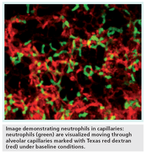
A new imaging technique has allowed researchers to observe the live interaction of living cells and the immune response to lung injury
In a recent study, performed at the University of California, San Francisco, CA, USA, researchers have developed a new technique to image a breathing lung in a live mouse. The method allows the observation of the living cells, and their interactions, and the immune response to lung injury, without disrupting the normal functioning of the organ.
Imaging of moving objects is difficult, and often results in blurred images; however, the researchers developed a custom rig, which was combined with super-fast imaging to achieve detailed images of the lung. “We figured out a method for holding cells still long enough to image them without interrupting their normal processes. This enabled us to observe cellular events as they happen naturally rather than the usual way, which is to stop the motion of cellular processes in order to photograph them,” explains Max Krummel, the senior author of the article.
The custom device applies a small amount of suction to the lung tissue surface, holding the tissue within the range of a two-photon microscope. An image of the tissue is then taken using super-fast imaging. The full biological process of different cells can be monitored by recording images 30 times per second.
Two-photon microscopy is a light-based high-resolution image technique that uses infrared pulsed lasers to penetrate tissue layers. The technology is capable of capturing small details, as small as one micron in diameter. The custom-built microscopes used in the study to carry out these measurements were built on site at the University of California, San Francisco.
The lung is a particularly difficult organ to image, due to the constant movement of the organ. However, the lung is the site of many important biological functions and is important in the immune response of the body. The lung is often a main point of contact with the outside environment, in terms of the inhalation of pathogens, allergens, air and toxins.
Previously, the only method for observing cellular activity was to stop the processes in order to image the cells. This new method allows observation of cellular activity in real time, allowing cellular function to be determined and observation of immune response as it occurs.
It is hoped that the development of this technique will allow scientists to examine the physiological aspects of disease and function of cells in more detail. The team plans to further miniaturize the rig in order to image live tissue biopsies. Krummel suggests, “Many different specialities of clinical scientists and pathologists can use this imaging method to learn how disease progression unfolds, but miniaturizing the rig to image biopsied tissue would be a tremendous improvement … for instance, we can catch how cells interact with tumors and observe whether they promote growth or rejection, and whether medical therapy is working.”
Source: Looney MR, Thornton EE, Lamm WJ et al.: Stabilized imaging of immune surveillance in the mouse lung. Nat. Methods 8, 91–96 (2010).


