Case Blog - Interventional Cardiology (2011) Volume 3, Issue 3
Open-ended coronary circulation: innocent bystander or a road map for intervention?
- Corresponding Author:
- Mohit D Gupta
Department of Cardiology, GB Pant Hospital and associated Maulana Azad Medical College, New Delhi, India
Tel: +91 119 8101 21311
Fax: +91 114 709 8755
E-mail: drmohitgupta@yahoo.com
Abstract
Keywords
coronary cascade,coronary cascade,intercoronary communication,open-ended circulation
Interarterial communication has been described in some regions of the human arterial system, such as the Circle of Willis and the intestinal branches of the superior mesenteric artery [1]. However, intercoronary communication (ICC) is a rare condition in which there is open-ended circulation with uni- or bi-directional blood flow between two coronary arteries [1]. Only a few cases of ICC have been reported since its first description [2–6]. The clinical significance of these communications has remained uncertain. In this article, we report a series of cases from our center describing various types of ICC. We also report two newer types of communications and highlight the clinical relevance of this unique entity for the cardiologist in light of the information in the existing literature and the present series.
Case reports
▪ Case 1
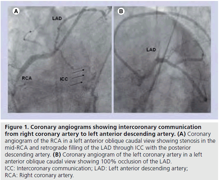
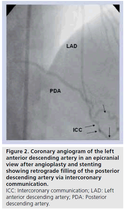
A 45‑year-old male, who is a known case of coronary artery disease (CAD) with anterior wall myocardial infarction and who was on regular treatment, presented to our center with worsening angina. 2D echocardiogram revealed hypokinesia of the inferior wall with a global left ventricular (LV) ejection fraction of 45%. His coronary angiography showed occlusion of the proximal left anterior descending artery (LAD) and 90% stenosis in the mid-segment of the right coronary artery (RCA) (Figure 1A & 1B). Retrograde filling of the LAD occurred by an ICC from the posterior descending artery (PDA) of the RCA (Figure 1A & Supplementary Video 1, see online at www.futuremedicine. com/toc/ica/3/3). The patient underwent percutaneous coronary intervention (PCI) of the LAD followed by the RCA. The lesion in the LAD was crossed with Persuader 6 wire (Medtronic Vascular, CA, USA). Sequential dilatation with sprinter 1.5 × 6 mm (Medtronic Vascular) and 2 × 6 mm (Medtronic Vascular) balloons was administered. However, antegrade flow in the LAD was not evident. In fact, contrast injection showed a unique ‘milking effect’, as if some retrograde flow in the LAD was competing with the antegrade flow (Supplementary Video 2). The procedure was completed by stenting the LAD with two stents – ProNova 2.5 × 28 mm (Vascular Concepts, India) and ProLink 2.5 × 15 mm (Vascular Concepts). The final angiogram showed flow in ICC from the LAD to the RCA (Figure 2 & Supplementary Video 3). PCI of the RCA with one drug-eluting ProNova stent was carried out after 1 day. After completion of PCI of the RCA, it was noticed that the ICC was not visualized on injection from either the RCA (Supplementary Video 4) or the LAD. There were no other collateral vessels visible. This was our first encounter with this strange anomaly.
Figure 1: Coronary angiograms showing intercoronary communication from right coronary artery to left anterior descending artery. (A) Coronary angiogram of the RCA in a left anterior oblique caudal view showing stenosis in the mid-RCA and retrograde filling of the LAD through ICC with the posterior descending artery. (B) Coronary angiogram of the left coronary artery in a left anterior oblique caudal view showing 100% occlusion of the LAD. ICC: Intercoronary communication; LAD: Left anterior descending artery; RCA: Right coronary artery.
Figure 2: Coronary angiogram of the left anterior descending artery in an epicranial view after angioplasty and stenting showing retrograde filling of the posterior descending artery via intercoronary communication. ICC: Intercoronary communication; LAD: Left anterior descending artery; PDA: Posterior descending artery.
▪ Case 2
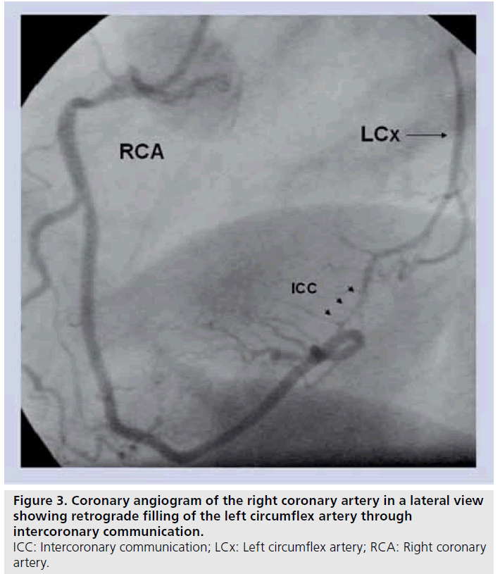
A 60‑year-old female with class II angina and an inconclusive exercise test underwent coronary angiography. A left system angiogram revealed normal epicardial arteries. Right coronary angiogram showed a normal RCA with visualization of the distal left circumflex artery (LCx) (Figure 3). A detailed examination of the results of the angiographic study identified a unidirectional intercoronary connection between the RCA and the LCx, which was formed by a single vessel that followed the course of the posterior atrioventricular groove (Figure 3).
Figure 3: Coronary angiogram of the right coronary artery in a lateral view showing retrograde filling of the left circumflex artery through intercoronary communication. ICC: Intercoronary communication; LCx: Left circumflex artery; RCA: Right coronary artery
▪ Case 3
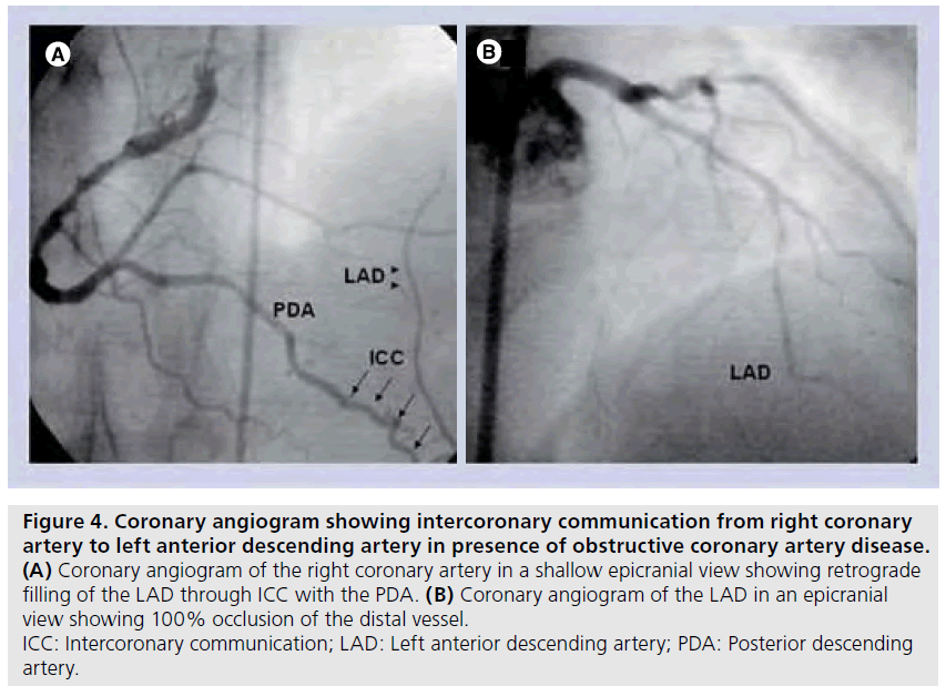
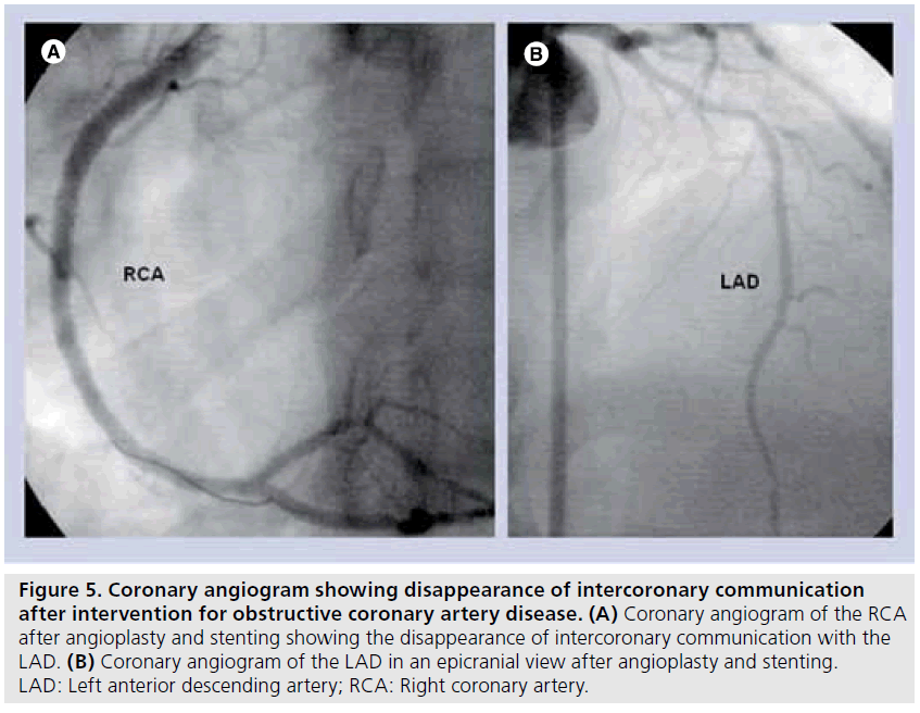
A 50‑year-old male, a known case of anterior wall myocardial infarction, presented with postmyocardial infarction angina. Echocardiogram showed no regional wall motion abnormality with a global ejection fraction of 65%. The RCA angiogram showed 80% stenosis in the proximal and mid-portion (Figure 4A). Left system angiogram showed complete occlusion of the distal LAD with retrograde filling up to the point of occlusion by an ICC from the PDA (Figure 4B). There were no other collateral vessels. After PCI of the LAD and RCA, ICC between the PDA and LAD disappeared (Figure 5A & 5B).
Figure 4: Coronary angiogram showing intercoronary communication from right coronary artery to left anterior descending artery in presence of obstructive coronary artery disease. (A) Coronary angiogram of the right coronary artery in a shallow epicranial view showing retrograde filling of the LAD through ICC with the PDA. (B) Coronary angiogram of the LAD in an epicranial view showing 100% occlusion of the distal vessel. ICC: Intercoronary communication; LAD: Left anterior descending artery; PDA: Posterior descending artery.
Figure 5: Coronary angiogram showing disappearance of intercoronary communication after intervention for obstructive coronary artery disease. (A) Coronary angiogram of the RCA after angioplasty and stenting showing the disappearance of intercoronary communication with the LAD. (B) Coronary angiogram of the LAD in an epicranial view after angioplasty and stenting. LAD: Left anterior descending artery; RCA: Right coronary artery.
▪ Case 4
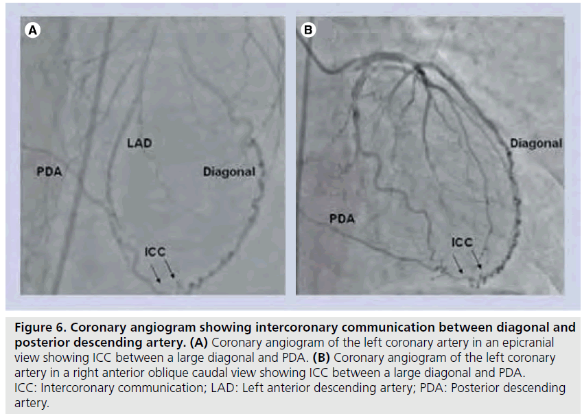
A 65‑year-old male, a known case of chronic stable angina with a positive treadmill test, underwent coronary angiography. This revealed an eccentric 75% lesion in the Ramus intermedius and a 100% occlusion of the ostial RCA. The left coronary angiogram identified type 1 LAD and an ICC between the large diagonal and the PDA of the RCA (Figure 6A & 6B).
Figure 6: Coronary angiogram showing intercoronary communication between diagonal and posterior descending artery. (A) Coronary angiogram of the left coronary artery in an epicranial view showing ICC between a large diagonal and PDA. (B) Coronary angiogram of the left coronary artery in a right anterior oblique caudal view showing ICC between a large diagonal and PDA. ICC: Intercoronary communication; LAD: Left anterior descending artery; PDA: Posterior descending artery.
▪ Case 5
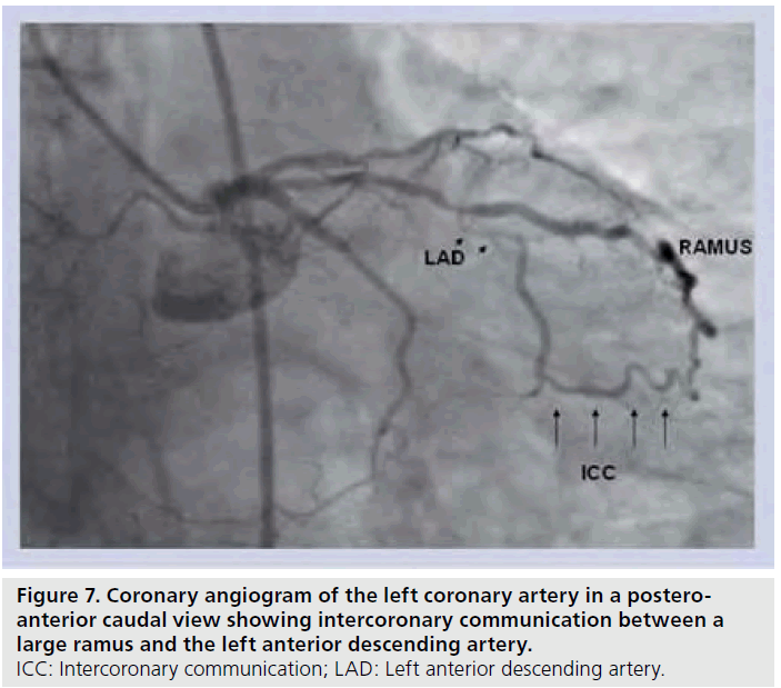
A 56‑year-old male, a known case of anterior wall myocardial infarction 2 years previously, presented with progressively worsening angina for the last 1 year. His global ejection fraction was 60%. Coronary arteriography showed occlusion of the LAD in the mid-segment with retrograde filling by an ICC arising from a large ramus (Figure 7 & Supplementary Video 5). No other collaterals were observed.
Figure 7: Coronary angiogram of the left coronary artery in a posteroanterior caudal view showing intercoronary communication between a large ramus and the left anterior descending artery. ICC: Intercoronary communication; LAD: Left anterior descending artery.
Discussion
Coronary cascade, better known as ICC or open-ended coronary circulation, is rare [1]. It is defined as an open-ended circulation with uni- or bi-directional blood flow between two coronary arteries [2,4]. The true prevalence of ICC is unknown and has been estimated to be approximately 2.3 per 100,000 population [7]. Two types of ICC have been defined: communication between the LCx and RCA in the posterior atrioventricular groove (as in case 2) and communication between the LAD and PDA in the distal interventricular groove (as in cases 1 and 3). The former type is more common [7]. In this article, we describe two newer types of ICC: between the large diagonal and PDA (as in case 4) and between the ramus and LAD (as1 in case 5). These communications have never been reported previously.
Intercoronary communication is distinct from the collateral arteries as they are larger in caliber (≥1 mm), extramural and follow a rectilinear epicardial course [1,7,8,9]. On the other hand, vessels belonging to the collateral circulation are usually tortuous, smaller and generally multiple in number. However, septal collateral circulation may not be tortuous. Also, the histologic structure of the connecting vessel in ICC has the characteristic of a normal arterial wall with a well-defined muscular layout [8]. It is suggested that faulty embryological development allows the existing intercoronary channel to remain prominent and maintain a large caliber [4]. On the other hand, collateral channels develop later as a result of ischemic stimulation. Although coronary artery flow in ICC is more commonly unidirectional, and sometimes bidirectional, there is always flow from the right to left coronary artery in the absence of underlying CAD [9]. However, this may not be the case in the presence of significant obstruction in the RCA.
The hemodynamic nature of collateral circulation and ICC has not been studied systematically. This may be of importance as it may provide exact functional differentiation between these two entities. Based on anatomical characteristics of ICC, it may be hypothesized that the hemodynamic measurements in these channels might be the same as that of the native vessel. However, this may not be the case in collateral circulation. In the present series, these measurements were not performed as our aim was to emphasize the existence of this underrecognized entity. However, study of hemodynamic parameters in future reports will further establish the role of ICC. There are conflicting views regarding the clinical and functional implications of ICC. Almost all patients with ICC with normal coronary arteries have underlying chest pain [1–3]. The mechanism of this pain is not clear. It has been hypothesized that it could be due to myocardial ischemia, if the ICC causes a coronary steal phenomenon resulting in inadequate perfusion [7]. Most of the reported cases of ICC did not have underlying obstructive CAD [1,2,9]. However, four out of five cases in the present series had underlying severe obstructive CAD. It has also been proposed that ICC may play a protective role if a lesion develops in one of the two vessels it links together. Evidence for this is conflicting [10,11]. However, in the present series, all cases with ICC and underlying obstructive CAD had relatively preserved LV systolic function despite having severe underlying CAD (as in cases 1 and 3). These cases demonstrate that ICC may have a protective role if an obstructive CAD develops in any of the communicating arteries. We believe that these channels provide more blood flow than collaterals to the obstructed artery due to their larger caliber. Hence, in their presence, LV systolic functions may be relatively preserved, as in the present series. The larger caliber of these communications probably also explains the unique milking effect due to competitive flow observed in case 1. Such a competitive flow is never seen with the collateral circulation, as these channels are less than 200 p in diameter and are generally not as effective as ICC in providing blood flow to the occluded artery [8].
Intercoronary communication may also serve as a better guide in opening the occluded artery, for two reasons. First, owing to its anatomic connection with the main vessel, it will always provide an exact guide to the lumen of the artery being opened in an antegrade approach. Second, owing to its larger caliber and histological features similar to the normal arterial wall, it may serve as a good channel for retrograde approach to opening of the occluded artery if the antegrade approach fails.
Disappearance of ICC after PCI of both the vessels (cases 1 and 3) is a unique feature. It shows that anatomical ICC may be present in many cases but angiographic visualization is only seen in a few. In a postmortem study of 100 human hearts from individuals with an average age of 61 years, seven cases had ICC [11]. This shows a relatively higher anatomical prevalence of approximately 7%, compared with a much lower angiographic prevalence of 0.002%. This also suggests that these channels may remain dormant in undiseased vessels and assume clinical significance in the presence of obstructive CAD. The authors also believe that the reported low angiographic prevalence of this entity is because of under-recognition, which in turn may be due to unawareness of this unique anomaly.
The present case series provides an insight into these enigmatic connections. It suggests that open-ended circulation is not just a benign observation, but may have a protective role if obstructive CAD develops. Furthermore, it may act as a guide for an interventional cardiologist in opening the occluded artery. Awareness of these communications may be useful to a cardiologist.
Future perspective
Intercoronary communication is an extremely rare condition in which there is an open-ended circulation with uni- or bi-directional blood flow between two coronary arteries in the presence or absence of obstructive CAD. They are distinct from collateral coronary circulation. Hemodynamic pressure measurements in ICC may help in further characterization of these unique communications and to help distinguish them from collaterals. The presence of ICC is of distinct advantage both to the patient as well as the cardiologist. Owing to their larger caliber, they are able to provide more complete blood flow to the jeopardized area than a coronary collateral, which is of smaller caliber. This results in relatively preserved LV function even in the presence of a completely occluded vessel. Functional measurements in these channels will further characterize them from collateral circulation. For an aggressive interventional cardiologist, these channels can serve as a channel for a retrograde approach to opening the occluded artery. Awareness of this entity will help in prompt recognition of these channels, which otherwise largely remain undiagnosed or are confused with a normal collateral.
Financial & competing interests disclosure
The authors have no relevant affiliations or financial involvement with any organization or entity with a financial interest in or financial conflict with the subject matter or materials discussed in the manuscript. This includes employment, consultancies, honoraria, stock ownership or options, expert testimony, grants or patents received or pending, or royalties.
No writing assistance was utilized in the production of this manuscript.
Executive summary
Intercoronary communications are extremely rare
▪▪ Intercoronary communications are an open-ended circulation with uni- or bi-directional blood flow between two coronary arteries. They may occur in the absence or presence of obstructive coronary artery disease.
Intercoronary communications are distinct from coronary collaterals
▪▪ Intercoronary communications are larger than coronary collaterals, extramural and follow a rectilinear epicardial course. They have the same histological structure as a normal vessel. They are caused by faulty embryological development, which leads to persistent intercoronary channels.
Intercoronary communications provide more complete blood supply to the occluded artery
▪▪ Owing to the larger caliber compared with the collateral circulation, intercoronary communication provides much better circulation when present in the presence of an occluded artery.
Intercoronary communications may be helpful in intervention
▪▪ Intercoronary communication may serve as a perfect guide to an interventional cardiologist when opening the occluded artery in an antegrade fashion. They may also serve as a potential route for retrograde opening of the occluded vessel.
References
Papers of special note have been highlighted as:
▪ of interest
▪▪ of considerable interest
- Kursaklioglu H, Barcin C, Iyisoy A: Intercoronary communication with unidirectional blood flow. J. Invasive Cardiol. 16(5), 269–270 (2004).
- Cheng TO: Arteriographic demonstration of intercoronary arterial anastomosis in a living man without coronary artery disease. Angiology 23, 76–88 (1972).
- Dubel HP, Gliech V, Rutsch W: Communication between nonstenotic coronary arteries. Z. Kardiiol. 92, 273–275 (2003).
- Atak R, Guray U, Akin Y: Images in cardiology: intercoronary communication between the circumflex and right coronary arteries distinct from coronary collaterals. Heart 88, 29 (2002).
- Carangal VP, Dehmer GJ: Intercoronary communication between the circumflex and right coronary arteries. Clin. Cardiol. 23, 125–126 (2000).
- Meireles GC, Abreu Filho LM: Intercoronary connection with bidirectional blood flow and concentric left ventricular hypertrophy. Rev.Paul. Med. 112, 654–657 (1994).
- Sokmen A, Tuncer C, Sokmen G, Akcay A, Koroglu S: Intercoronary communication between the circumflex and right coronary arteries: a very rare coronary anomaly. Hellenic J. Cardiol. 50, 66–67 (2009).
- Fournier JA, Cortacero JAPF, de la Llera LD, Sánchez A, Arana E, Morán JE: Distal intercoronary communication. A case report and medical literature review. Rev. Esp. Cardiol. 56(10), 1026–1028 (2003).
- Gavrielatos G, Letsas KP, Pappas LK et al.: Open ended circulation pattern: a rare case of a protective coronary artery variation and review of the literature. Int. J. Cardiol. 112, e63–e65 (2006).
- Donaldson RF, Isner JF: Intercoronary continuity: an anatomic basis for bidirectional coronary blood flow distinct from coronary collaterals. Am. J. Cardiol. 53, 351–352 (1984).
- Arat-Ozkan A, Gurmen T, Ersanli M, Okçün B, Babalik E, Küçükoglu MS: A patient with bicuspid aorta and intercoronary continuity: a rare variant of coronary circulation. Jpn Heart J. 45, 153–155 (2004).
▪ Discusses intercoronary communication (ICC) between the right coronary artery and the distal left circumflex artery and discusses the possible significance of these communications.
Discusses ICC between the right coronary artery and the distal left circumflex artery and also highlights differences between collateral circulation and ICCs.
▪▪ Describes existing types of ICC and how they differ from normal collateral circulation.
