Clinical images - Imaging in Medicine (2017) Volume 9, Issue 2
Optic nerve transection after blunt facial trauma
L Turco*1 & B Phillips21Department of Surgery, University of Kansas Medical Center, Kansas City, Kansas, USA
2Department of Surgery & Department of Clinical Science and Translational Research, Creighton University School of Medicine, Omaha, Nebraska, USA
- Corresponding Author:
- L Turco
Department of Surgery
University of Kansas Medical Center
Kansas City, Kansas, USA
E-mail: LTurco@kumc.edu
Abstract
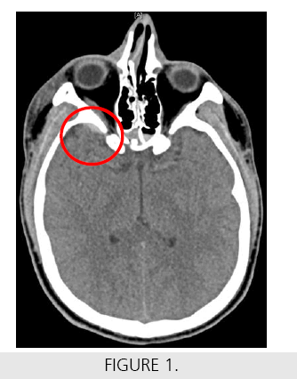
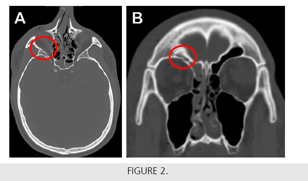
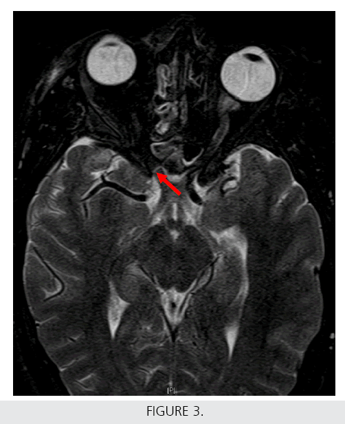
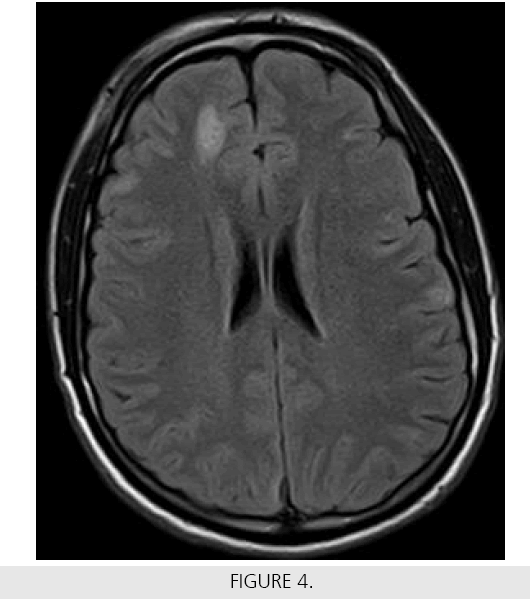
A 22-year-old male was transferred by EMS from an outside hospital to our Level I Trauma Center after being found in a pool of blood 11 feet away from a 7 foot wall. The exact mechanism of his injury was unknown. On presentation to our center, the patient was intubated and sedated. He was noted to have a right periorbital laceration and a splint on his right forearm. Radiographs from the outside hospital demonstrated distal right radial and ulnar fractures. Computed tomography (CT) imaging revealed scattered areas of subarachnoid hemorrhage in the superior right and left frontal lobe (Figure 1) as well as numerous right-sided facial fractures involving the lateral maxillary wall, zygomatic arch, lateral pterygoid plate, greater and lesser wing of the sphenoid, and the superior orbital rim (Figures 2A and 2B). The patient was admitted to the ICU where he remained confused, but tolerated extubation. Shortly after being transferred to a general medical ward, he complained of complete loss of vision in his right eye. A contrastenhanced MRI was acquired and T2-weighted images revealed a transverse hyperintensity and enhancement (arrow in Figure 3) along the proximal intracanalicular portion of the right optic nerve suggestive of right optic nerve transection. FLAIR MRI showed residual cerebral intraparenchymal hemorrhage along the right parafalcine frontal lobe (Figure 4). The patient was discharged home and seen by an outpatient ophthalmologist who reported that his vision loss is permanent. He continues to follow-up with occupational therapy and in our outpatient trauma clinic.


