Clinical images - Imaging in Medicine (2017) Volume 9, Issue 6
Osteolytic bone lesions: a rare complication of myelofibrosis
Zeineb Alaya*, Abderrahim Khlif & Elyes BouajinaFarhat Hached Hospital Sousse, Tunisia
- Corresponding Author:
- Zeineb Alaya
Farhat Hached Hospital Sousse, Tunisia
E-mail: zeineb_a@hotmail.fr
Abstract
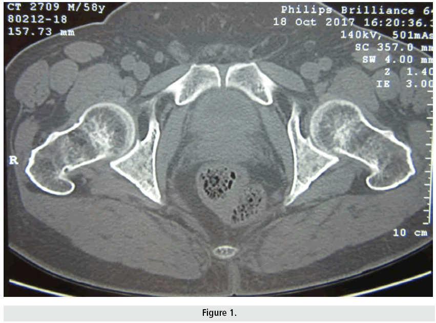
A 62 year old man was referred to our clinic because of inflammatory pelvic bone pain that has been evolving for 3 months. The patient has been followed in hematology since 2005 for chronic myeloid splenomegaly. Pelvic X-ray was normal. MRI of the pelvis showed a heterogeneous aspect of the bone matrix. The CT of the pelvis objectified pathological bone matrix with lytic multigeodic appearance due to his hematopathy (FIGURE 1). Biology showed hypercalcemia at 2.8 mmol/l. The osteolytic bone lesions presented by our patient are a rare complication of Myeloid Splenomegaly. The patient was treated with pamidronate.


