Case Report - International Journal of Clinical Rheumatology (2021) Volume 16, Issue 7
Rare cause of pigmenturia from anti-hydroxy-3-methylglutarylcoenzyme; A reductase (HMGCR) induced muscle necrosis treated with immunosuppression: A Case Report and literature review
- *Corresponding Author:
- Ejo John
Platinum Hospitalist Group, Valley Health System, Las vegas, USA
E-mail: ejomjohn123@gmail.com
Abstract
Myositis and muscle necrosis can be associated with anti-HMGCR antibody in patients with prior HMG-CoA reductase exposure. There are isolated reports of muscle necrosis in patients with no prior history of statin exposure. We percent a case of severe muscle necrosis with no predisposing factors. Our patient had high anti-HMGCR antibody titers causing rhabdomyolysis, muscle necrosis requiring long-term immunosuppressive agents
Keywords
pigmenturia • muscle necrosis • immunosuppression
Introduction
Necrotizing myositis is a rare phenomenon usually associated with medications, trauma, infection, auto-immune disease, and electrolyte disturbance. We present a case of anti- HMGCR myositis without the forementioned causes.
Case presentation
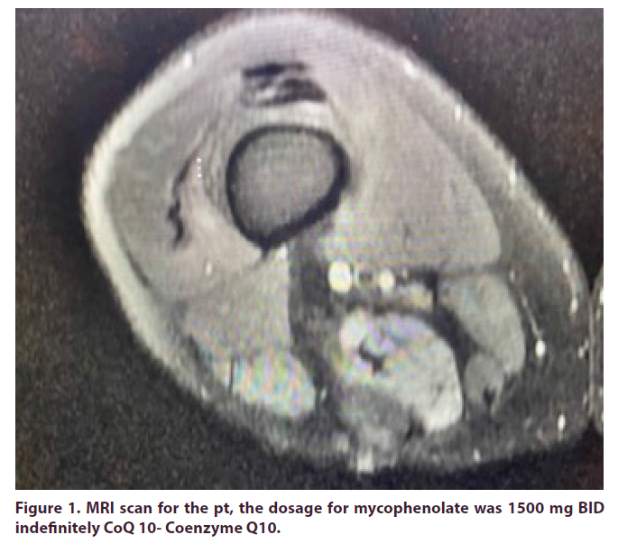
A 65-year-old woman with no past medical history presented to the hospital with generalized weakness and myalgias, predominantly in the lower extremity muscle group. She had no rash, fever, night sweats or chills. The patient did not take any medications at home. Her temperature was 36.9°C, heart rate 117 beats/minute and a blood pressure of 164/90 mmHg. Physical exam revealed right lower extremity strength 2/5 and left lower extremity strength 3/5. No other neurological deficits. Laboratory testing showed a creatinine kinase of 31,960 ng/mL, myoglobin level of 10,534 ng/mL with a urinalysis revealing hematuria but no RBC’s per high power field on microscopy. Immunological testing with anti-Jo 1, anti- PL-7, anti-PL-12, anti-EJ, anti-OJ, anti-SRP, antimitochondrial, anti-TIF-1, anti-MDA, anti-Jo 1, anti-SSA, SSB. Rheumatoid factor, SLE antibodies were all unremarkable. MRI of her lower extremity revealed evidence of extensive myositis. The patient underwent right vastus lateralis muscle biopsy which showed necrotic myofibers consistent with necrotizing myositis. There was no evidence of active inflammatory myopathy, vasculitis, congenital myopathy, muscular dystrophy, lipid storage myopathy or mitochondrial myopathy. Further serological testing revealed anti-HMGCR antibody ratio of>200 with a normal reference range of<20 with a strong positive result of greater than >60. The patient was diagnosed with anti-HMGCR myositis. The patient was started on prednisone and mycophenolate mofetil (Figure 1).
Discussion
Antibodies are usually produced to disrupt foreign pathogens. In the setting of immune mediated HMGCR antibodies, the body produces antibodies to destroy normal functioning HMG-CoA reductase. HMG-CoA reductase is a precursor for cholesterol synthesis. The precise mechanism of myopathy associated with its deficiency is unclear. It is hypothesized that the reduction in this enzyme prevents synthesis of mevalonic acid which is required to produce Coenzyme Q 10. With a reduction in Coenzyme Q 10 this can cause abnormal mitochondrial function leading to impaired energy production and subsequent myopathy [1].
This clinical syndrome is typically seen in patients with statin use. However, 33% of patients who have necrotizing myopathy from anti-HMGCR antibody have not been exposed to statin therapy. Patients who have not been exposed tend to have a higher creatinine kinase level than statin associated muscle necrosis [2].
Myoglobin is a heme containing protein and is released from muscular injury. However, this marker is rapidly metabolized to bilirubin and may not be apparent in the serum as it may return to normal levels within eight hours. Usually, creatinine kinase is elevated in the absence of myoglobin and typically rises within the first 12 h following injury and does not decline until three days post muscular injury. Both myoglobin and hemoglobin can be detected on urinalysis by dipstick, however without the presence of RBCs on high power field, this is usually indicative of pigmentary due to myoglobin [3, 4].
Immunosuppression is the mainstay treatment of immune mediated anti-HMGCR myositis with steroids as first line therapy. Other immunosuppressive agents such as azathioprine, methotrexate, mycophenolate mofetil, and rituximab have been tried to varying and inconclusive response. The duration of therapy is unknown but long-term immunosuppression is hypothesized.
Conclusion
Our patient was initiated on prednisone in conjunction with mycophenolate mofetil. With immunosuppression therapy, clinical symptoms improved, and creatinine kinase level returned to within normal limits. On repeat urinalysis, hematuria cleared. The patient was maintained on prednisone for one month followed by taper. Mycophenolate mofetil is currently being used as life-long immunosuppression maintenance therapy in our patient.
References
- Borch X, Poch E, Grau JM. Rhabdomyolysis ans acute kidney injury. N. Engl. JmMed. 361, 62–72 (2009).
- Mammen AL. Immune mediated necrotizing myopathy. Arthritis. Rheum. 62, 2757–2766 (2010).
- Mammen Al. Autoantibodies against HMG CoA reductase in patients with statin exposure. Arthritis. Rheum. 63, 713– 721 (2011).
- Werner Jl, Chritopher– Stine L, Ghazarian Sr et al. Antibodies level and correlation with creatine kinase. Arthritis. Rheum. 64, 4087–4093 (2012).


