Clinical images - Imaging in Medicine (2017) Volume 9, Issue 3
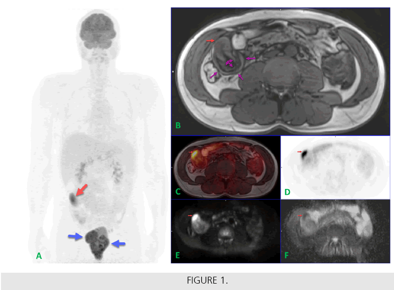
Relapsing lymphoma provoking small intestinal intussusception assessed with PET/MR
Zoltán Tóth*, Peter Zadori, Gabor Lukacs, Peter Rajnics, Miklos Egyed, Imre Repa & Arpad KovacsMedicopus Non-profit Ltd. Health Centre, University of Kaposvár, Kaposvar, Hungary
- Corresponding Author:
- Zoltán Tóth
Medicopus Non-profit Ltd. Health Centre
University of Kaposvár, Kaposvar, Hungary
E-mail: toth.zoltan@ke.hu
Abstract
A 37 year old male patient was referred to 18F-fluorodeoxyglucose (FDG) PET/MR examination due to suggested pelvic diffuse large B-cell lymphoma (DLBCL) relapse. Anterior aspect Maximal Intensity Projection (MIP) PET image (FIGURE A) visualized radiotracer accumulating pelvic recurrences (blue ararows) and focal FDG uptake in the right abdominal area (red arrow). Identical to the circumscribed right abdominal radiopharmacon accumulation on the PET scan (FIGURE A and D; red arrow) the T1- weighted opposed-phase axial DIXON MR image revealed wall thickening along the terminal ileum (FIGURE B, red arrow) with high signal intensity area on the diffusion weighted MR sequence (FIGURE E; red arrow) and diffusion restriction on the corresponding ADC map (FIGURE F; red arrow). The FDGaccumulating intestinal wall thickening can be observed on the fused PET/MR image dataset (FIGURE C, red arrow) as well. In a close proximity ileoileal intussusception was also detected on the T1-weighted MR image (FIGURE B, purple arrows). As obstructive intestinal symptoms were developed, small bowel resection was carried out and pathology confirmed DLBCL relapse as a root cause of invagination.


