Clinical images - Imaging in Medicine (2020) Volume 12, Issue 4
Residual Cystic Stump Stones Following Cholecystectomie
Wael Ferjaoui*, Wafa Ghariani, Mohamed Ali Chaouch, Mehdi Khalfallah, Hichem Jerraya & Ramzi NouiraDepartment of surgery, Charles Nicolle Hospital, Tunis, Tunisia
- Corresponding Author:
- Wael Ferjaoui
Department of surgery
Charles Nicolle Hospital, Tunis, Tunisia
E-mail: Farjaouiwael4@gmail.com
Abstract
Keywords
Cholecystectomie ■ Cystic stump stones
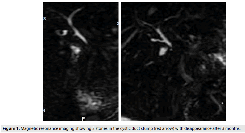
We present the case of a 63 yr old female patient. Past medical history was unremarkable. She was operated 5 days ago for cholecystectomy. Currently, she presented with persistent abdominal pain in the right upper quadrant region,. Physical examination showed tenderness of the abdomen. Blood tests were normal. Magnetic resonance imaging was done. It showed that within the distal cystic duct stump, there was 3 rounded filling defects showing low attenuation that was highly concerning for residual cystic duct stone. A second check made by magnetic resonance imaging after 3 months confirmed the disappearance of these stones (FIGURE 1).
Residual cystic stump stones after a cholecystectomy remain a rare complication [1]. It is part of post cholecystectomy syndrome. Abdominal ultrasound and/or computed tomography often allow a first diagnostic approach.
In case of failure of the endoscopic treatment, surgical treatment by laparascopic is indicated and cholangio-MRI constitutes the reference preoperative examination [2]. A therapeutic abstention can be proposed since a spontaneous passage of stones in the main bile duct is possible
References
- Vyas FL, Nayak S, Perakath B et al. Gallbladder remnant and cystic duct stump calculus as a cause of postcholecystectomy syndrome. Trop. Gastroenterol. 26, 159-160, (2005).
- Mergener K, Clavien PA, Branch MS et al. A stone in a grossly dilated cystic duct stump: a rare cause of post cholecystectomy pain. Am. J. Gastroenterol. 94, 229-231, (1999).


