Clinical images - Imaging in Medicine (2017) Volume 9, Issue 6
Significantly enlarged ventricles due to ventriculoperitoneal shunt malfunction
Xiaoliang (Shawn) Qiu* & Fuad M ZeidDepartment of Internal Medicine, Marshall University, USA
- Corresponding Author:
- Xiaoliang (Shawn) Qiu
Department of Internal Medicine
Marshall University, USA
E-mail: qiuxi@marshall.edu
Abstract
Introduction
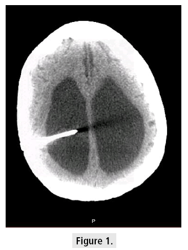
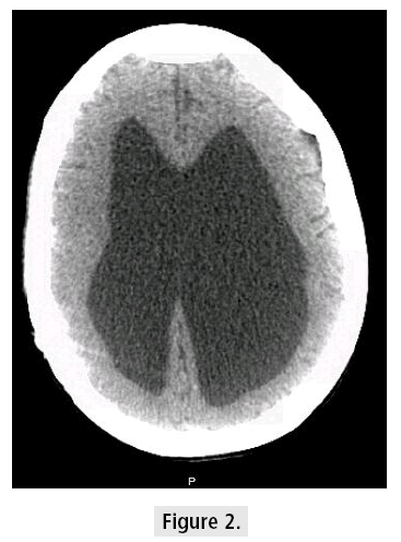
A 55 year old female with a past medical history significant for seizure, spina bifida status post ventriculoperitoneal (VP) shunt, and chronic respiratory failure with chronic tracheostomy on home trilogy ventilator presented with seizurelike activity and dysphagia. Patient was not on any antiepileptic drugs and she did not have seizure for a few years. Electroencephalography revealed no electrographic seizures or interictal epileptiform activity. Head CT showed significant dilatation of the lateral and third ventricles (FIGURES 1 and 2). Further history revealed she was initially shunted at birth and has had only 3 revisions since then. The current shunt in place was done at the age of 11. It was not clear why no further shunt revision was done. However, most recent CT head from 2011 showed similar findings with no obvious interval changes. VP shunt placement is still the mainstay treatment for pediatric hydrocephalus. These devices have a relatively high complication and failure rate, often requiring multiple revisions, therefore such patients should receive regular follow-up through the transition to adulthood [1].


