Clinical images - Imaging in Medicine (2020) Volume 12, Issue 5
Spontaneous Isolated Superior Mesenteric Artery Dissection Mimics Acute Enteritis
Shao Hsuan Weng*
- Corresponding Author:
- Shao Hsuan Weng
Department of Radiology
Taipei Medical University Shuang-Ho Hospital
New Taipei City, Taiwan
Email: dororo320001@gmail.com
Abstract
Keywords
Acute enteritis ■ SISMAD
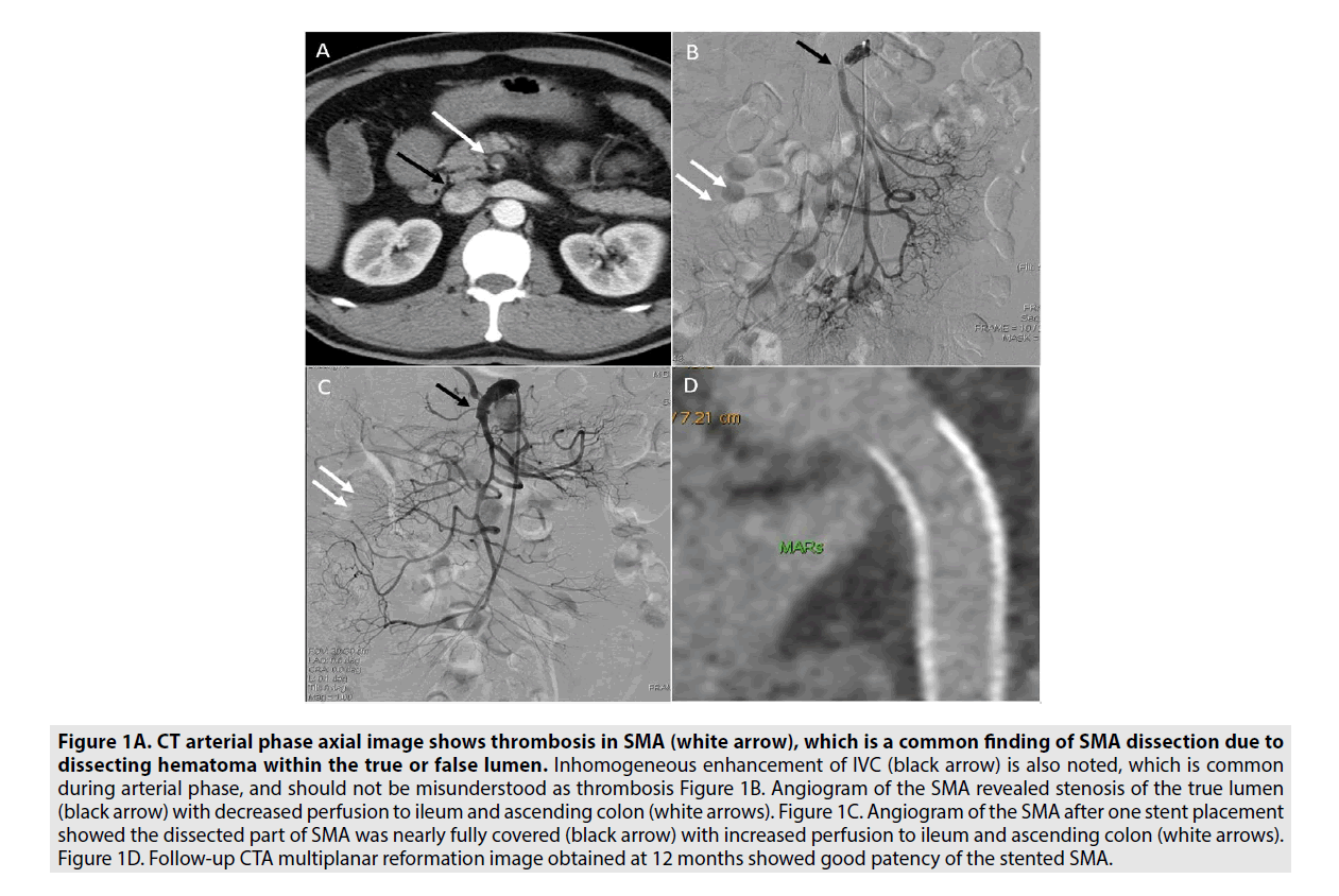
A 51yr old man presented to the emergency department with lower abdomen pain and vomiting for the last 2 days. There was no preceding history of trauma, surgery. Physical examination revealed lower abdomen tenderness. Mild decreased bowel sound. Laboratory data, ECG, chest X-ray were normal. He had been discharged with a diagnosis of enteritis. However, due to persistent abdomen pain, he re-attended and abdomen CT was obtained. The abdomen CT showed thrombosis in Superior Mesenteric Artery (SMA), which is a common finding of SMA dissection due to dissecting hematoma within the true or false lumen (FIGURE 1A). Inhomogeneous enhancement of IVC is also noted, which is common during arterial phase, and should not be misunderstood as thrombosis (FIGURE 1A).
Figure 1A. CT arterial phase axial image shows thrombosis in SMA (white arrow), which is a common finding of SMA dissection due to dissecting hematoma within the true or false lumen. Inhomogeneous enhancement of IVC (black arrow) is also noted, which is common during arterial phase, and should not be misunderstood as thrombosis Figure 1B. Angiogram of the SMA revealed stenosis of the true lumen (black arrow) with decreased perfusion to ileum and ascending colon (white arrows). Figure 1C. Angiogram of the SMA after one stent placement showed the dissected part of SMA was nearly fully covered (black arrow) with increased perfusion to ileum and ascending colon (white arrows). Figure 1D. Follow-up CTA multiplanar reformation image obtained at 12 months showed good patency of the stented SMA.
Followed angiogram revealed stenosis of the true lumen of SMA with decreased perfusion to ileum and ascending colon, consistent with SMA dissection (FIGURE 1B).
After a stent was placed, the dissected part of SMA was nearly fully covered with increased perfusion to bowel loops (FIGURE 1C). The patient was uneventful thereafter. Follow-up CTA multiplanar reformation image obtained at 12 months showed good patency of the stented SMA. (FIGURE 1D).
Spontaneous Isolated Superior Mesenteric Artery Dissection (SISMAD) is a rare disease with emergency. Clinical symptoms may present with nonspecific or acute abdominal pain, cold sweating, which mimics enteritis.2 Clinical history and laboratory data are often unhelpful. In patients complicated with persistent pain, bowel ischemia, or aneurysm formation, stent placement or surgical revascularization is indicated.3 And stent placement is preferable due to its minimal invasiveness. Early and accurate detection is important to save life and avoid severe consequence [1-3].
References
- Luan J, Li XJ. Surgery E. Computed tomography imaging features and classification of isolated dissection of the superior mesenteric artery. Eur. J. Vasc. Endovasc. Surg. 46, 232-235, (2013).
- Min SI, Yoon KC, Min SK et al. Current strategy for the treatment of symptomatic spontaneous isolated dissection of superior mesenteric artery. Circulation. 54, 461-466, (2011).
- Kim J, Yoon CJ, Seong N et al. Spontaneous Dissection of Superior Mesenteric Artery: Long-Term Outcome of Stent Placement. J. Vasc. Interv. Radiol. 28, 1722-1726, (2017).


