Clinical images - Imaging in Medicine (2017) Volume 9, Issue 3
Sudden deafness due to lipiodol retention: unusual high density nonmetallic findings on CT
Sergio Ghirardo*Department of Medical and Surgical Sciences Health, University of Trieste, Trieste, Italy
- *Corresponding Author:
- Sergio Ghirardo
Department of Medical and Surgical Sciences
Health University of Trieste, Trieste, Italy
E-mail: ghirardo.sergio@gmail.com
Abstract
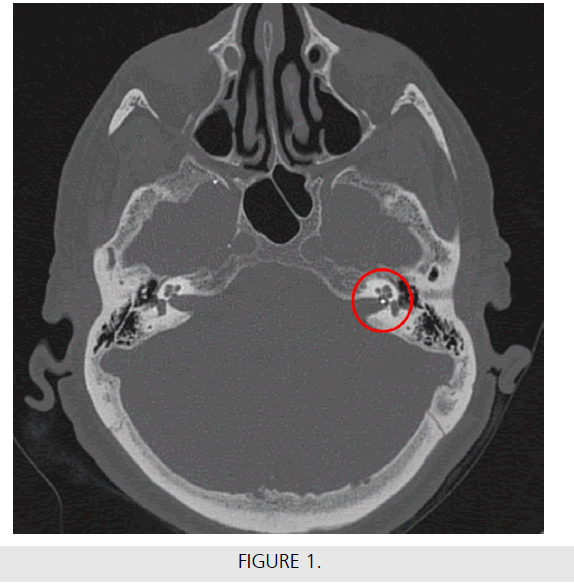
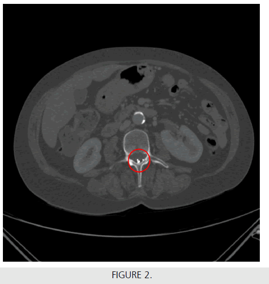
A 63 years old woman presents to otorhinolaryngologist with sudden unilateral sensorineural hearing loss and underwent a CT scan of the head, which ruled out a cerebral infarction. However, CT revealed five high density (nearly 380 Hounsfield units; more than the iron) spherical objects in the subarachnoid space, one of which located at the emerging of the left acoustic nerve from the petrous, and compressing it as shown in figure (FIGURE 1). Other radiological findings were compatible with the age of the patient. A previous abdomen CT scans had shown similar findings in lumbar vertebral canal, in subarachnoid space which had not been evaluated (FIGURE 2). Her clinical history was negative for trauma or surgery, but was notable for chemoembolization of a hepatocellular carcinoma, 6 years before. This extremely dense object is compatible with Lipiodol retention, a rare complication of such intervention [1]. On the other hand, localizations in the subarachnoid space are typical of a myelography [2], but there was no evidence of such intervention in the patient history. A neurosurgical consultation was performed, but the object was too close to the nerve to be removed. Patient adapted to hear with the contralateral ear and by now, she has not developed any other symptoms. A radiological follow up was considered, but in absence of an available therapy we preferred a wait and see approach.


