Case Report - Interventional Cardiology (2020) Volume 12, Issue 3
Transcatheter ASD closure in a patient with Dextroversion: A case report
- Corresponding Author:
- Yasmin Abdelrazek Ali
Department of Cardiology
Ain Shams University, Cairo, Egypt
E-mail: yasminabdelrazek@med.asu. edu.eg
Abstract
Background: Cardiac dextroversion is location of the heart in the right chest with the left ventricle remaining in the normal position to the left but lying anterior to the right ventricle. To the best of our knowledge no specific technique has been described before for successful transcatheter secundum atrial septal defect closure in patients with dextroversion.
Case presentation: We present a case of secundum atrial ASD in a patient with dextroversion, situssolitus, AV concordance and VA concordance. Patient was referred for transcatheter closure of ASD. Her transthoracic echocardiography showed a 7 mm secundum ASD, Upper normal RV size, small restrictive VSD 2 mm and a dilated main pulmonary artery. Transesophagealechocardiography showed an 11 mm defect with abnormal orientation of inter atrial septum due to cardiac dextroversion. Usual technique for positioning of ASD Amplatzer device (ASO 11) failed with prolapsing of the device into right atrium. Failed right upper pulmonary vein technique. With successful positioning of the device across the interatrial septum using left upper pulmonary vein technique.
Conclusion: We recommend left upper pulmonary vein technique for safe and direct positioning of ASD device across interatrial septum in patients with dextroversion.
Keywords
Congenital heart disease; Left upper pulmonary vein; Dextrocardia
Abbreviations
2D: 2 Dimensional; ASD: Atrial Septal Defect; ASO: Atrial Septal Occluder; AV: Atrioventricular; CXR: Chest X-ray; ECG: Electrocardiography; IAS: Interatrial Septum; LUPV: Left Upper Pulmonary Vein; MSCT: Multislice Computed Tomography; PA: Pulmonary Artery; PR: Pulmonary Regurgitation; RA: Right Atrium; RUPV: Right Upper Pulmonary Vein; RV: Right Ventricle; TEE: Transesophageal Echocardiography; VA: Ventriculoarterial; VSD: Ventricular Septal Defect
Background
Dextroversion of the heart is a type of dextrocardias, the heart is located in the right hemithorax without inversion of chambers. This congenital anomaly is the result of a counter-clockwise rotation of a normally developed heart in the right hemi thorax. It may be associated with different cardiac and pulmonary anomalies, or may be isolated. It has been described under different names: dextrorotation [1], isolated dextrocardia [2], dextrocardia with atria in situs solitus [3].
In 1998 Hakim et al., reported a case of ASD transcatheter closure in a case of dextrocardia with situsin versus, they used left atrium angiogram in the right anterior oblique view with cranial angulation to visualize the IAS, followed by successful positioning of Amplatzer device across the defect [4].
Maldjian et al., studied a case of isolated dextroversion by MSCT in 2006 and described that the right atrium was posterior and to the right of the left atrium. The morphologic right ventricle was posterior, slightly superior and to the right of the morphologic left ventricle [5].
In dextroversion, the interatrial septum is directed to the right, with the morphologic right atrium situated to the right and slightly posteriorly, and the morphologic left atrium to the left and slightly anteriorly. Galal et al., reported successful closure of fenestrated septum by Amplatzer cribriform device in a case with situs solitus and dextrocardia, they reported difficulty in device positioning, yet with no specific technique recommended [6].
Case Presentation
We present a case of a female patient 3-year-old, 11 Kg. She is the 2nd in order of birth of 2 children of non-consanguineous marriage. Her mother complained of recurrent chest infection. She had irrelevant perinatal history and irrelevant family history. On examination, the maximum impulse was felt to the right of the sternum, with no precordial bulge, no thrill, and harsh pansystolic murmur heard to the right of the sternum.
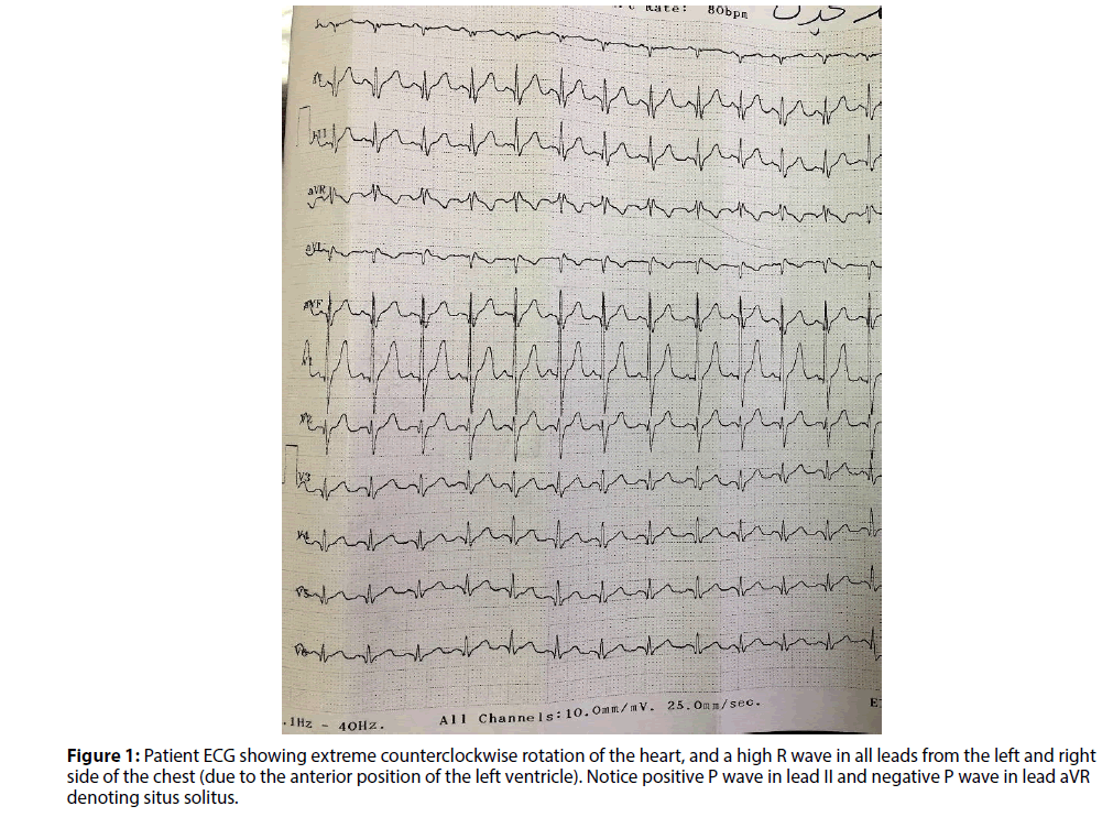
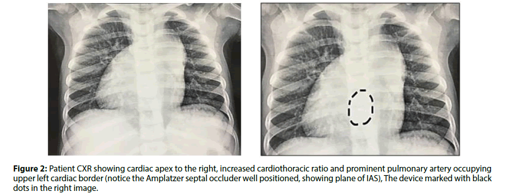
ECG showed extreme counterclockwise rotation of the heart, and a high R wave in all leads from the left and right side of the chest (due to the anterior position of the left ventricle) (Figure 1). CXR showed cardiac apex to the right with prominent pulmonary artery occupying upper left cardiac border and prominent pulmonary vascular markings till the outer 1/3 of pulmonary tree (Figure 2).
Transthoracic echocardiography showed visceral and cardiac situssolitus, AV concordance, VA concordance, secundum ASD measuring 7 mm shunting left to right with adequate rims all around except for absent aortic rim. Small perimembranoussubaorticVSD (ventricular septal defect) 2 mm shunting restrictively left to right. Upper normal RV size with dilated main pulmonary artery.Mild PR (pulmonary regurgitation) with estimated normal mean pulmonary artery pressure.
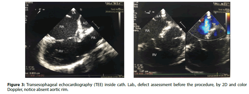
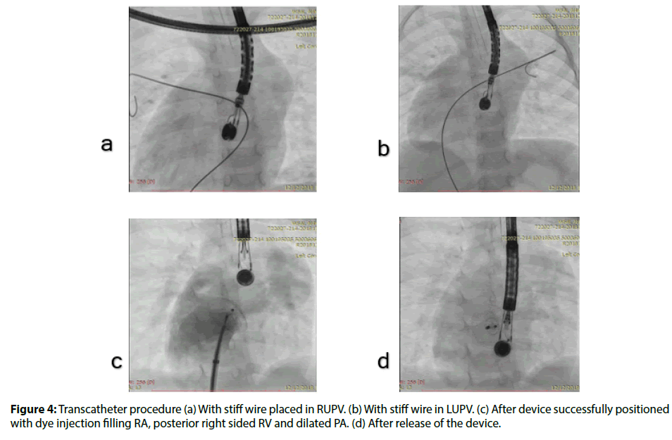
Cardiac catheterization under general anesthesia was done. We noticed marked rotation of the ventricles by the right turn of the catheter from the right atrium into the right ventricle, and by the right to the left direction of the main pulmonary artery. Transesophageal echocardiography was used to guide transcatheter ASD closure, which showed a secundum ASD measuring 11 mm in its maximum diameter, with adequate rims all around except for absent aortic rim.Normal pulmonary venous drainage into left atrium and normal systemic venous drainage to right atrium were assured (Figure 3). An Amplatzer septal occluder 11 was advanced across long sheath with left atrial disk opened in left atrium. Multiple trials were done to place the device across the IAS but failed with device prolapse into RA (right atrium). One trial to place the device from the right upper pulmonary vein was done but failed due to device prolapsing into RA as well. Finally, successful placement of the device was achieved through a left upper pulmonary vein technique by release of left atrial disc into left upper pulmonary vein followed by release of right atrial disc and withdrawal of the device to be properly positioned across the IAS (Figures 4-6 and Videos 1 and 2).
Figure 3: Transesophageal echocardiography (TEE) inside cath. Lab., defect assessment before the procedure, by 2D and color Doppler, notice absent aortic rim.
Figure 6: Transthoracic echocardiography 3 months after the procedure with well seated device in both (a) Subcostal view and (b) Apical 4 chamber view. Notice abnormal orientation of the septal plane.
Post procedure ECG showed normal sinus rhythm with no heart block. CXR showed well seated device (Figure 2). The patient was discharged home on Aspirin and instructions for endocarditis prophylaxis for 6 months. Follow up transthoracic echocardiography 3 months after the procedure showed well seated device with no residual flow across (Figure 6 and Videos 3-5)
Conclusion
To the best of our knowledge no specific technique has been described before for successful transcatheter secundum ASD closure in patients with dextroversion. We recommend left upper pulmonary vein technique for safe and direct positioning of ASD device across interatrial septum in patients with dextroversion.
References
- Ayres SM, Steinberg I. Dextrorotation of the heart. Circulation. 27(2): 268-274 (1963).
- Shaher RM, Johnson AM. Isolated laevocardia and isolated dextrocardia: pathology and pathogenesis 1963. Guy's Hospital Reports. II2: I27 (1963).
- Calder6n J, Marquez J, Cerezo L, et al. Dextrocardias con auriculas 'in situ solito'. Revista Espaniol ade Cardiologia. I8: 40 (1965).
- Hakim F, Madani A, Samara Y, et al. Transcatheter Closure of Secundum Atrial Septal Defect in a Patient with Dextrocardia Using the Amplatzer Septal Occluder. Cathet Cardiovasc Diagn. 43: 291–294 (1998)
- Maldjian PD, Saric M, Anis A. CT appearance of isolated dextroversion. Int J Cardiovasc Imaging. 22: 731-733 (2006).
- Galal MO, Khan MA, El-Segaier M. Percutaneous closure of atrial septal defect with situs solitus and dextrocardia. Asian Cardiovascular & Thoracic Annals. 23(2) 202-205 (2015).


