Case Report - International Journal of Clinical Rheumatology (2020) Volume 15, Issue 6
Transient Osteoporosis of hip during pregnancy
- Corresponding Author:
- Kawtar Nassar
Department of Rheumatology, Faculty of Medicine
Ibn Rochd University Hospital, Morocco
E-mail: nassarkawtar@gmail.com
Abstract
Transient osteoporosis of pregnancy is a rare condition that causes temporary bone loss. The syndrome is characterized by self-limited course and spontaneous resolution after 6 to 12 months. Several pathogenesis hypotheses have been proposed. Clinical manifestations include sudden onset of pain. MRI is considered the best diagnostic test for this condition in regard to sensitivity and specificity and in monitoring of disease progression. We report a 38-year-old pregnant woman presented during the third trimester of her pregnancy with right hip pain that became progressively severe. Imaging of her bilateral hips with MRI demonstrated increased signal in the bone-marrow of the right femur head and neck with minimal effusion. Repeat MRI performed at five months postpartum revealed resolution of edema.
Keywords
transient osteoporosis • pregnancy • hip • magnetic resonance imaging
Introduction
Transient osteoporosis of pregnancy is a rare, nondestructive and self-limiting condition with spontaneous resolution after 6 to 12 months [1]. The hip is the most frequent disease site. The diagnosis is often difficult in the early stages, due to lack of awareness and suspicion by the treating clinicians. It could be a subset of Complex Regional Pain Syndrom, as it has many similarities with it, namely, unknown cause, characteristic pain [2]. MRI is considered the best diagnostic test for this condition in regard to sensitivity and specificity and in monitoring of disease progression [3]. An early and accurate diagnosis can lead to early resolution of symptoms of the patients and avoid unnecessary investigations and treatment for other mimicking conditions. This condition is usually be managed by conservative and symptomatic means. We report a case of 38-year-old pregnant woman presented during the third trimester of her pregnancy with right hip pain secondary to transient osteoporosis.
Case report
The patient, F.A, 38-year-old G1P1, without past pathological history. She consulted for mechanical right hip pain during the ninth month of pregnancy that progressively worsened with time without any other symptoms. The physical exam found pain in hip mobility, especially in the abduction movement and external rotation, without joints limitation. There was no other clinical abnormally.
The biological test in particular; blood count, sedimentation rate and C-reactive protein was normal. The calcium rate was normal at 91 mg/l and the 25(OH) vitamin D2-3 was decreased at 24 ng/ml.
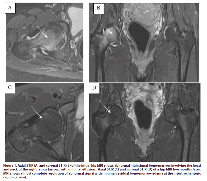
The Magnetic Resonance Imaging (MRI) of the hips was performed while the patient was in the third trimester of pregnancy. She revealed increased signal in the bone-marrow of the right femur head and neck with minimal effusion (Figure 1A and 1B). The patient was treated by physical therapy and was recommended to rest and use a 4-point walker. The evolution was favorable since a week of treatment.
Figure 1: Axial STIR (A) and coronal STIR (B) of the initial hip MRI shows abnormal high signal bone-marrow involving the head and neck of the right femur (arrow) with minimal effusion. Axial STIR (C) and coronal STIR (D) of a hip MRI five months later. MRI shows almost complete resolution of abnormal signal with minimal residual bone-morrow edema at the intertrochanteric region (arrow).
According to the literature result, findings were most characteristic of transient osteoporosis in pregnancy. A repeat MRI performed five months postpartum showed almost complete resolution of abnormal signal with minimal residual bone-morrow edema at the intertrochanteric region (Figure 1C and 1D). At the time of the second MRI, the patient’s symptoms had markedly improved and the physical exam was normal.
Discussion
Transient Osteoporosis of the Hip (TOH) is a poorly understood and forgotten clinical entity [1]. The diagnosis is often delayed. It was reported first by Ravault (1947) followed by Curtiss and Kincaid in 1959 [2] and has been described with different names such as Bone Marrow Edema (BME) syndrome, transient demineralization, complex regional pain syndrome type 1, migratory osteolysis, and algodystrophy of the hip [3]. It’s more frequently in pregnant women in their third trimester 1 [4]. The TOH is a nondestructive and self-limiting condition of the hip, which responds well to the conservative treatment. The TOH could be a subset of complex regional pain syndrome type 1 [5], as it has many similarities in clinical presentation and management. The TOH is associated with reduced mobility of the hip without a specific laboratory finding. The etiopathogenesis of TOH may include microvascular injury, transient ischemic episode, nontraumatic reflex sympathetic dystrophy, metabolic, and endocrine factors. A neurogenic hypothesis is advocated a, child's head compressing mother's obturator nerve [6,7]. MRI is considered the best diagnostic test for this condition in regard to sensitivity and specificity. She also helps in ruling out other pathologies [8-10]. Patients have often pain-free within an average of 2.8 weeks and had no limp in their gait. The pre-treatment and post-treatment MRI show resolution of bone morrow edema on week 12 in approximately 84% of patients (16/19) after 3 months of treatment [9].
A symptomatic and supportive treatment is recommended with protected weight bearing and graduated physiotherapy regime. An average time of 7.5 months (range, 4-11 months) was required for complete bone-morrow edema resolution on MRI and an average time of 5.8 months (range, 2-10 months) for resolution of clinical symptoms [10,11]. An early and accurate diagnosis can lead to early resolution of symptoms of the patients. This condition is usually be managed by conservative and symptomatic means [12].
Conclusion
We report a 38-year-old pregnant woman presented during the third trimester of her pregnancy with right hip pain that became progressively severe. Imaging of her bilateral hips with MRI demonstrated increased signal in the bone-marrow of the right femur head and neck with minimal effusion. Repeat MRI performed at five months postpartum revealed resolution of edema.
Compliance with ethical standards
This study did not require any funding.
Conflicts of interest
All the authors are no conflicts of interest related to this manuscript. Kawtar Nassar is the patient’s attending physician. She made the bibliography research and she wrote the manuscript. Saadia Janani participated at the bibliography research, she read and approved the manuscript.
Ethical approval
This article does not contain any studies with human participants or animals performed by any of the authors. This is a case report, and informed consent was obtained from the patient included in the study.
References
- Patel V, Temkin S, O'Loughlin M. Transient osteoporosis of pregnancy in a 32-year-old female. Radiol. Case. Rep. 7(2), 646 (2011).
- Curtiss PH, Kincaid WE. Transitory demineralization of the hip in pregnancy. A report of three cases. J. Bone. Joint. Surg. Am. 41-A, 1327–1333 (1959).
- Vande Berg BC, Lecouvet FE, Koutaissoff S et al. Bone marrow edema of the femoral head and transient osteoporosis of the hip. Eur. J. Radiol. 67, 68-77 (2008).
- Hadidy AM, Al Ryalat NT, Hadidi ST et al. Male transient hip osteoporosis: Are physicians at a higher risk? Arch. Osteoporos. 4(1-2), 41–45 (2009).
- Shaik Ahmed, Jeanine M. Lindsay, RN BSN et al. Spinal Cord Stimulation for Complex Regional Pain Syndrome: A Case Study of a Pregnant Female. Pain. Physician. 19(3), 487-493 (2016).
- Trevisan C, Klumpp R, Compagnoni R. Risk factors in transient osteoporosis: A retrospective study on 23 cases. Clin. Rheumatol. 35(10), 2517–2522 (2016).
- Dunstan CR, Evans RA, Somers NM. Bone death in transient regional osteoporosis. Bone. 13, 161–165 (1992).
- Dawid Szwedowski EF, Zaneta Nitek EF, Jerzy Walecki EF. Evaluation of transient osteoporosis of the hip in magnetic resonance imaging. Polish. J. Radiol. 79, 36–38 (2014).
- Hofmann S, Engel A, Neuhold A et al. Bone-marrow oedema syndrome and transient osteoporosis of the hip. An MRI-controlled study of treatment by core decompression. J. Bone. Joint. Surg. Br. 75(2), 210–216 (1993).
- Balakrishnan A, Schemitsch EH, Pearce D et al. Distinguishing transient osteoporosis of the hip from avascular necrosis. Can. J. Surg. 46(3), 187–192 (2003).
- March RM, Tovaglia V, Meo A et al. Transient osteoporosis of the hip. Hip. Int. 20, 297–300 (2010).
- Agarwala S, Vijayvargiya M, Single Dose Therapy of Zoledronic Acid for the Treatment of Transient Osteoporosis of Hip. Ann. Rehabil. Med. 43(3), 314–320 (2019).


