Image Article - Imaging in Medicine (2021) Volume 13, Issue 9
Unexpected concomitant cervical and endometrial cancer revealed by 18F-FDG PET-CT
- Corresponding Author: E-mail: salah.nabihoueriagli@gmail.com
Abstract
Case Description
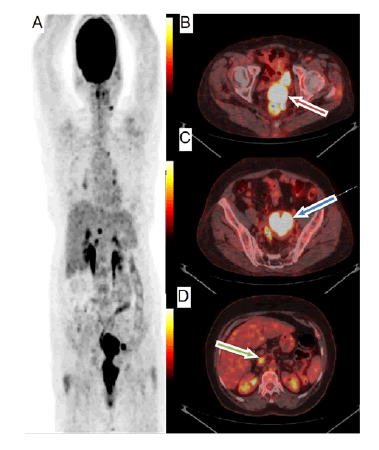
A 65-years-old post-menopausal female patient, presented with menorrhagia and pelvic pain. Vaginal examination finds a suspicious cervical mass. Biopsy confirmed the diagnosis of cervix adenocarcinoma and CT showed external left iliac lymphadenopathy. 18F-FDG PETCT was performed to assess the expansion evaluation, and uncovered notwithstanding the hypermetabolism of the cervix (SUVmax=24.4), a significant hypermetabolism in the uterine cavity (SUVmax=30.1) which can't be identified with a straightforward intra uterine dying. We do speculate a myometrial attack, however correlative MRI shows discontinunity between the two neurotic cycles. Our investigation prompts perform hysteroscopy, and biopsy of uterine divider uncovered an accompanying endometrial malignant growth. We do likewise report in our 18F-FDG PET-CT nodal expansion in the outside left iliac, predominant mesenteric and meso-rectum regions (FIGURE 1).
Figure 1: (A) Maximum Intensity Projection (MIP), showing in addition to the pathological cervix hypermetabolism and the lomboarortic lymphadenopathy involvement, an important uptake in the uterine cavity related to the concomitant endometrial cancer. (B) Fusion image in axial section showing intense hyper metabolism in cervix (SUVmax=24.4) (Red arrow). (C) Fusion image in axial section showing intense uptake in the uterine cavity (SUVmax=30.1) (Blue arrow). (D) Fusion image in axial section showing intense uptake in lomboarortic lymphadenopathy (SUVmax=7.9) (Green arrow).
In women’s cancers, all imaging studies (CT, MRI &18F-FDG PET-CT) evaluate the presence of lymph nodal involvement, and detect local and distant metastatic disease at initial diagnosis [1,2]. 18F-FDG PET-CT is recommended to evaluate lomboaortic lymphadenopathy involvement in the context of loco regional extension with 86% of sensitivity and 94% of specificity. In the uterine and endometrial cancers, this exploration is indicated respectively in stages IB2 and II FIGO [3]. 18F-FDG PETCT detects myometrial extension from the cervical stroma which can alter management [4].
References
- Grigsby PW, Siegel BA, Dehdashti F. Lymph node staging by positron emission tomography in patients with carcinoma of the cervix. J Clin Oncol. 19(17), 3745-3749 (2001).
- Yen TC, Lai CH, Ma SY, et al. Comparative benefits and limitations of 18F-FDG PET and CT-MRI in documented or suspected recurrent cervical cancer. Eur J Nucl Med Mol Imaging. 33(12), 1399-1407 (2006).
- Standards, Options et Recommandations. Utilisation de la tomographie par émission de positons au [18F]-FDG en cancérologie. Bull Cancer 90, S51- S52 (2003).
- Chou HH, Chang TC, Yen TC, et al. Low value of [18F]-fluoro-2-deoxy-D-glucose positron emission tomography in primary staging of early-stage cervical cancer before radical hysterectomy. J Clin Oncol. 24(1), 123-128 (2006).


