Case Report - Imaging in Medicine (2020) Volume 12, Issue 5
Vertebral Fracture Mimicking Infectious Spondulodiscitis
Soumaya Boussaid*, Yasmine Makhlouf, Samia Jemmali, Sonia Rekik & Mohamed ElleuchRheumatology department, La rabta Hospital, Tunis, Tunisia
- Corresponding Author:
- Soumaya Boussaid
Rheumatology department
La rabta Hospital, Tunis, Tunisia
E-mail: soumayaboussaid@hotmail.com
Abstract
A 73yr old man presented to the rheumatologic departement of our institute with inflammatory neck pain evolving since 3 years that has gradually worsened six months ago . He reported an alteration of the general condition . A cervical X ray as well as medullary MRI were performed. Imaging features may be consistent with spondylodiscitis (SD). However the patient’s clinical presentation, the absence of an infectious etiology, MRI data supported the presumptive diagnosis of Fractured vertebrae on a degenerative cervical myelopathy . The patient was immobilized with a cervical collar. He received intravenous Dexamethasone (4mg) during a week followed by physiotherapy sessions.
Keywords
pondylodiscitis vertebral fracture ■ COPD ■ Mimicking
Introduction
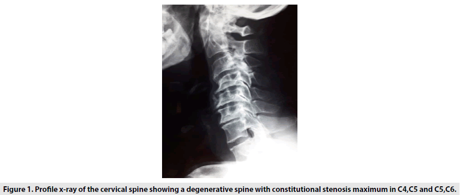
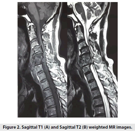
A 73yr old man with a history of chronic obstructive pulmonary disease (COPD), smoking the amount of at least 60 pack-years of cigarettes /cessation of 3 years, presented to the rheumatologic departement of our institute with inflammatory neck pain evolving since 3 years that has gradually worsened six months ago . He reported an alteration of the general condition , anorexia and a weight loss of 15 kilos in few months as well as a recent history of dysphonia . Systemic examination was unremarkable. There was no fever. Neck movement was limited in all axes and there was a muscular stiffening but no neck swelling or crepitus were found. Neurological examination didn’t show abnormalities . Otorhinological examination revealed a polyp of the vocal cord. The white blood cell count (WBC), the Erythrocyte Sedimentation Rate (ESR), and the C-reactive Protein (CRP) level were normal. A direct laryngoscopy under General Anesthesia was performed to remove the polyp. Twentyfour hour after the intervention , the patient developed a brutal tetraplegia with a urinary incontinence . Neurological examination noted a quadripyramidal syndrome. A cervical X ray (FIGURE 1) as well as medullary MRI were performed (FIGURE 2).
Figure 1: Profile x-ray of the cervical spine showing a degenerative spine with constitutional stenosis maximum in C4,C5 and C5,C6.
Diagnosis
Fractured vertebrae C5 on a degenerative cervical myelopathy. A cervical spine MRI showed hyper signal in the cervical cord on T2-weighted sagittal section , hyper signal of the C4,C5,C6 cervical vertebrae thickening of the posterior longitudinal ligament. cervical spinal stenosis; multilevel degenerative changes in the cervical spine which was consistent with the signal intensities of intervertebral discs are slightly decreased.
Commentary
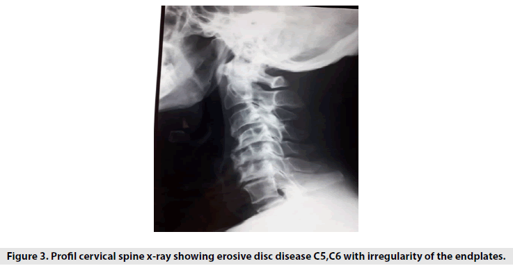
Imaging features may be consistent with spondylodiscitis (SD). However the patient’s clinical presentation, the absence of an infectious etiology, MRI data supported the presumptive diagnosis of Fractured vertebrae on a degenerative cervical myelopathy . The patient was immobilized with a cervical collar. He received intravenous Dexamethasone (4mg) during a week followed by physiotherapy sessions. . At follow up, the patient responded well to treatment with an Improvement of sphincter control and a complete motor function recovery. A lateral cervical spine x-ray was performed after 3 months showing erosive disc disease C5,C6 with irregularity of the endplates. (FIGURE 3)
Discussion
Spinal vertebral fractures are common in elderly patient especially those who suffer from a degenerative cervical myelopathy (DCM). Indeed, DCM may result in static and dynamic injury to the spinal cord. Left untreated, it can lead to tetraplegia and wheelchair dependence. Neck tenderness or neurologic deficits even after a minor trauma should increase the suspicion of cervical spinal fracture [1]. The diagnosis requires confirmation via imaging and MRI is the preferred modality [2]. Our case was a very confusing and unusual case of fractured vertebrae that mimicked findings of infectious spondylodiscitis on imaging. MRI is a powerful tool to diagnose spondylodiscitis. Indeed, MRI may show bone marrow infiltrations in contiguous spinal segments with low signal intensity on T1-weighted image and high signal intensity on T2-weighted image of the vertebral body and endplate. In our case, Intervertebral discs and disc spaces were relatively spared, which is a possible finding in both DCM and spondylodiscitis. Moreover, Subligamentous spread of inflammatory soft tissue and multi-level contiguous or non-contiguous involvements are also seen in spondylodiscitis [3].
Besides, MRI findings of involvement of 3 contiguous vertebrae and paraspinal soft tissue infiltrative change with relatively preserved intervertebral discs may be more indicative of spinal metastasis than infectious spondylodiscitis [4]. Thus, the differential diagnosis may still be difficult in similar cases in future. A patient’s history of malignancy should also be considered in the differential diagnosis. A CT-guided biopsy should be performed for a histopathological confirmation [5].
Conclusion
Despite its likeness to infectious spondylodiscitis on MRI, other entities such as DCM that can mimick the radiographic findings of SD shoud be considered. History and clinical examination remain essential for diagnosis.
References
- Mohamed IN, Fatema EA, Nada Ahmed et al. Breast Cancer: Conventional Diagnosis and Treatment Modalities and Recent Patents and Technologies. Breast. Cancer. 9, 17-34, (2015).
- Malik SA, Murphy M, Connolly P et al. Evaluation of morbidity, mortality and outcome following cervical spine injuries in elderly patients. Eur. Spine. 17, 585-591, (2008).
- Harrop JS, Naroji S, Maltenfort M et al. Cervical myelopathy: a clinical and radiographic evaluation and correlation to cervical spondylotic myelopathy. Spine. 35, 620-624, (2010).
- Hong SH, Choi JY, Lee JW et al. MR imaging assessment of the spine: infection or an imitation? Radiographics. 29, 599-612, (2009).
- Lee CM, Lee S, Bae J. Contiguous Spinal Metastasis Mimicking Infectious Spondylodiscitis. J. Korean. Soc. Radiol. 73, 408-412, (2015).


