Case Report - Interventional Cardiology (2019) Volume 11, Issue 5
When it pops-out: Restart all over again from the beginning?
- Corresponding Author:
- Takehiro Yamashita
Department of Cardiology, Cardiovascular Center, Hokkaido Ohno Memorial Hospital, Sapporo, Hokkaido, Japan E-mail: t_yamashita@cvc-ohno.or.jp
Received date: August , 2019; Accepted date: August 26, 2019; Published date: September 02, 2019
Abstract
An 82-year-old female with a severe aortic stenosis (AS) and a history of acute decompensated heart failure was scheduled for TAVR. A stiff wire accidentally dislodged from the left ventricle before delivering an S3 valve to the aortic valve plane. A successful S3 implantation was achieved by crossing a regular wire using the flex wheel of the delivery system handle to align the nose cone with the aortic orifice followed by an exchange to a stiff wire. This is the very first clinical case report of this technique for a possible bailout procedure during a premature LV guidewire dislodgement, which significantly reduces additional risk, procedural time and cost.
Keywords
Transcatheter aortic valve implantation/replacement (TAVI/TAVR); Complication; Guidewire dislodgment; Bailout
Case Description
The dislodgment of a stiff wire from the Left ventricular (LV) cavity when a Sapien valve is already in the patient’s vasculature is a rare but troublesome complication of transcatheter aortic valve replacement (TAVR). A theoretical bailout technique of retrying to cross the aortic valve with a regular wire using the original system has been suggested in a literature [1]. However, such clinical cases have never been reported. An 82-year-old female with a severe aortic stenosis (AS) and a history of acute decompensated heart failure was scheduled for TAVR [2-10].
Discussion and Conclusion
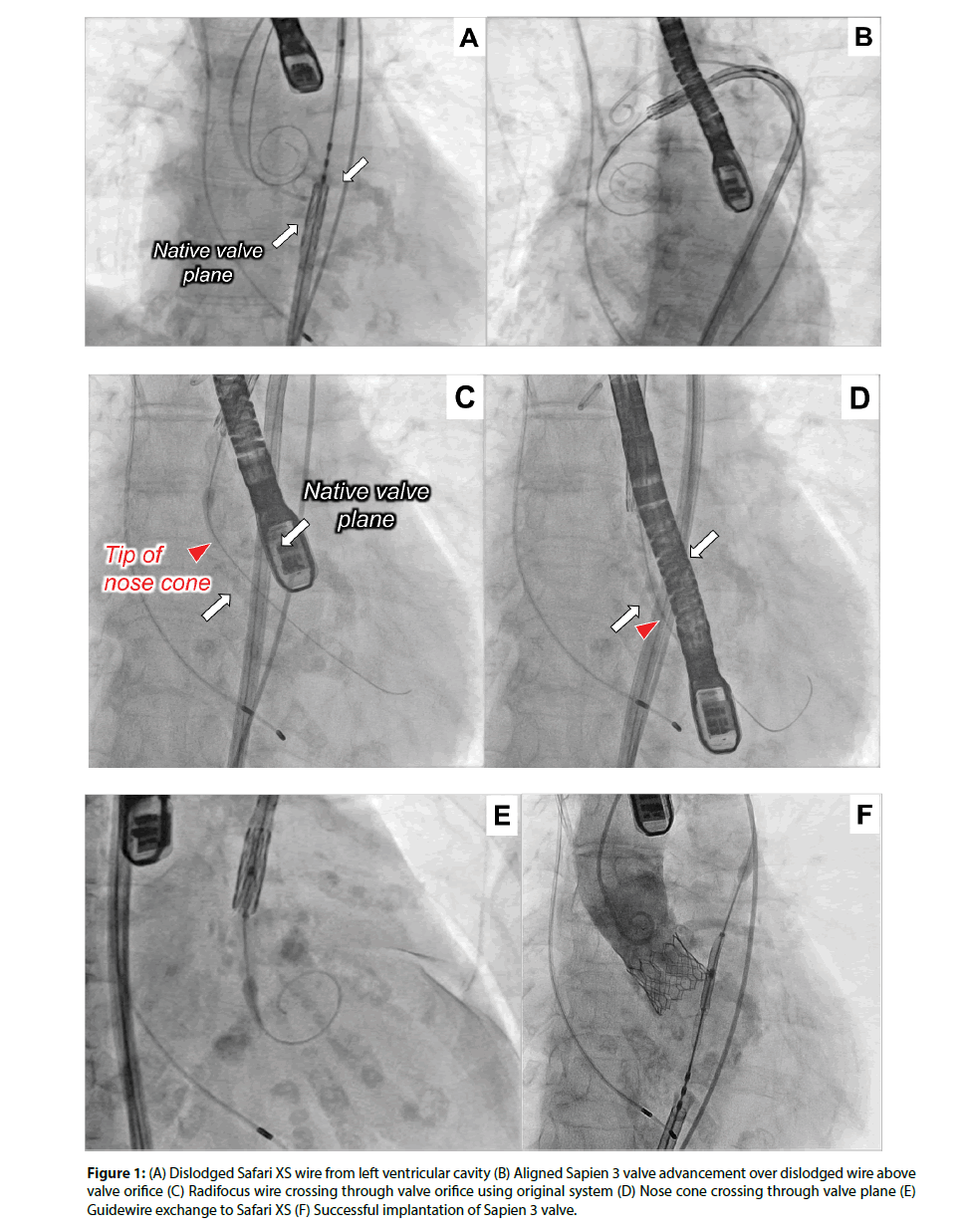
Pre-procedural echocardiography results indicated the stenosis was very severe with the measurements of aortic valve area (AVA) 0.28 sq cm, AVA index 0.22, mean pressure gradient 95.9 mmHg and peak velocity 6.1 m/sec. Predilatation was performed using a 20-mm balloon with a Safari XS guidewire (Boston Scientific, Boston, MA, USA) placed in the LV but the guidewire accidentally came out during our struggle to cross a crimped 23-mm Sapien 3 valve (Edwards Lifesciences, Irvine, CA, USA) inside an e-sheath through her right external iliac artery due to the extreme friction (Figure 1A). After successfully crossing the iliac artery, the valve was aligned to a proper position and delivered above her native aortic valve over the dislodged wire (Figure 1B). The Safari XS was then withdrawn and, using its flex function and manual torque of the entire system, the nose cone was manipulated in order to cross the valve orifice with a Radifocus wire (TERUMO, Tokyo, Japan). This guidewire was then successfully delivered into the LV (Figure 1C, Video 1) with just the tip of the nose cone going through the valve orifice (Figure 1D, Video 2), which enabled a guidewire exchange to a Safari XS (Figure 1E, Video 2) and the Sapien 3 implantation (Figure 1F). To the best of our knowledge, this is the very first clinical report using this technique as a bailout procedure during a premature LV guidewire dislodgement, which significantly reduced additional risk, procedural time and cost.
Figure 1: (A) Dislodged Safari XS wire from left ventricular cavity (B) Aligned Sapien 3 valve advancement over dislodged wire above valve orifice (C) Radifocus wire crossing through valve orifice using original system (D) Nose cone crossing through valve plane (E) Guidewire exchange to Safari XS (F) Successful implantation of Sapien 3 valve.
Relationship with Industry
Drs. Yamashita, Iwakiri, Kurebayashi, Suzuki and Ohkawa have reported no relationships relevant to the contents of this paper to disclose.
Supplementary Data
Fluoroscopic imaging of the technique. Using its flex function and manual torque of the entire system, the nose cone was manipulated in order to cross the valve orifice with a Radifocus wire. This guidewire was then successfully delivered into the LV (Video 1) with just the tip of the nose cone going through the valve orifice, which enabled a guidewire exchange to a Safari XS (Video 2).
References
- Dall’Alla G, Moretti C, Marrozzini C, et al. How should I treat an unexpected deadlock at the time of transcatheter aortic valve prosthesis implantation? Euro Intervention. 13: 256-258 (2017).
- Takahashi K, Domoto S, Isomura S, et al. Transcatheter aortic valve replacement for aortic stenosis with complicated anatomy. Report of a case. 72(9): 694-697 (2019).
- Cid-Menéndez A, López-Otero D, González-Ferreiro R, et al. Predictors and outcomes of heart failure after transcatheter aortic valve implantation using a self-expanding prosthesis. Rev Esp Cardiol (Engl Ed). pp: 1885-5857 (2019).
- Czerwińska-Jelonkiewicz K, Milewski K, Buszman P, et al. Peri-procedural hemostasis disorders in surgical and transcatheter aortic valve implantation. Postepy Kardiol Interwencyjnej. 15(2): 176-186 (2019).
- Scholtz S, Piper C, Horstkotte D, et al. Transcatheter Aortic Valve Implantation in Patients With Pre-Existing Mechanical Mitral Valve Prostheses. J Invasive Cardiol. 31(9): 260-264 (2019).
- Chiaroni PM, Ternacle J, Deux JF, et al. Computed tomography for transcatheter tricuspid valve development. Eur Radiol. p: 26 (2019).
- Ziccardi MR, Groves EM. Bioprosthetic valve fracture for valve-in-valve transcatheter aortic valve replacement: rationale, patient selection, technique, and outcomes. Interv Cardiol Clin. 8(4): 373-382 (2019).
- Dolci G, Vollema EM, van der Kley F, et al. One-year follow-up of conduction abnormalities after transcatheter aortic valve implantation with the sapien 3 valve. Am J Cardiol. pp: 30835-30855 (2019).
- Lader JM, Barbhaiya CR, Subnani K, et al. Factors predicting persistence of AV nodal block in post-TAVR patients following permanent pacemaker implantation. Pacing Clin Electrophysiol. 2019.
- Schneider AW, Hazekamp MG, Versteegh MIM, et al. Reinterventions after freestyle stentless aortic valve replacement: an assessment of procedural risks. Eur J Cardiothorac Surg. p: e22 (2019).


